

 |
 |
|
|
|
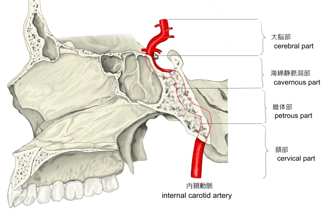
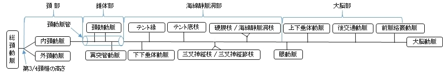
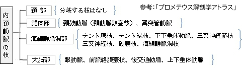
内頚動脈の区分と枝 |
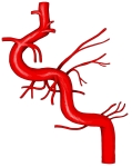
頭蓋における
内頚動脈の区分 |
|
|
|
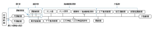

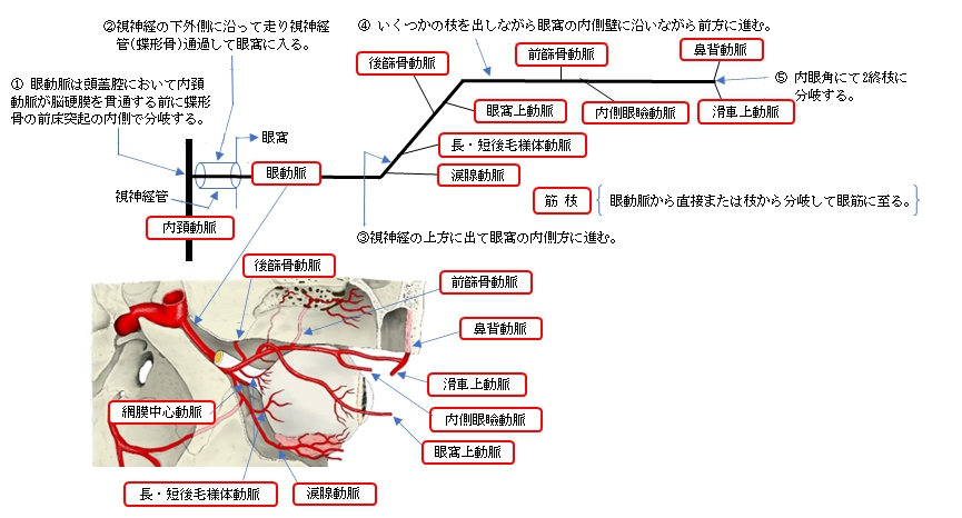
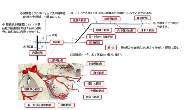
1. 頭蓋腔(内頚動脈が脳硬膜を貫く前)において蝶形骨の前床突起の内側で分岐する。
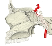
2. 視神経の下外側に沿って走り視神経管(蝶形骨)を通過して眼窩に入る。
3. 視神経の上に出て眼窩の内側方に進む。
4. いくつかの枝を出しながら眼窩の内側壁に沿いながらを前方に進む。
5. 内眼角に達して滑車上動脈と鼻背動脈の2終枝に分岐する。

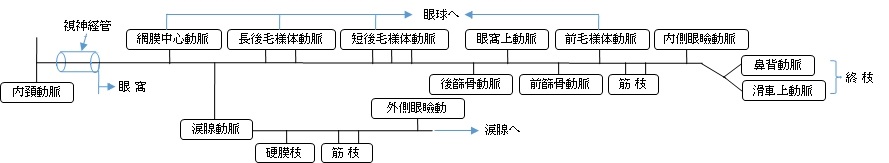
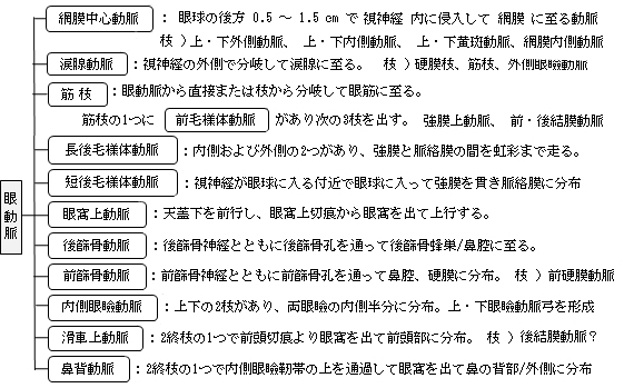
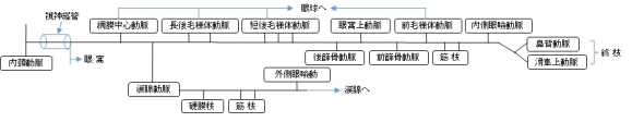
以下は「 日本人体解剖学 」を参考にして眼動脈の枝を簡単に一覧にしたものとなる。

以下は「 Wikipedia 」の解説文となる。
「 The ophthalmic artery (OA) is the first branch of the internal carotid artery distal to the cavernous sinus. Branches of the OA supply all the structures in the orbit as well as some structures in the nose, face and meninges. Occlusion of the OA or its branches can produce sight-threatening conditions.
【 structure 】
The OA emerges from the internal carotid artery usually just after the latter emerges from the cavernous sinus although in some cases, the OA branches just before the internal carotid exits the cavernous sinus. The OA arises from the internal carotid along the medial side of the anterior clinoid process and runs anteriorly passing through the optic canal with and inferolaterally to the optic nerve. The ophthalmic artery can also pass superiorly to the optic nerve in a minority of cases.[1] In the posterior third of the cone of the orbit, the ophthalmic artery turns sharply medially to run along the medial wall of the orbit. 」
【 語 句 】
・ distal : 末端の ・ cavernous sinus : 海綿静脈洞 ・ occlusion : 閉鎖 ・ anterior clinoid process : 前床突起
【 イラスト掲載サイト 】
・ イラストや写真を掲載しているサイト-Ⅰ
・ イラストや写真を掲載しているサイト-Ⅱ
・ イラストや写真を掲載しているサイト-Ⅲ
・ イラストや写真を掲載しているサイト-Ⅳ
・ イラストや写真を掲載しているサイト-Ⅴ

|