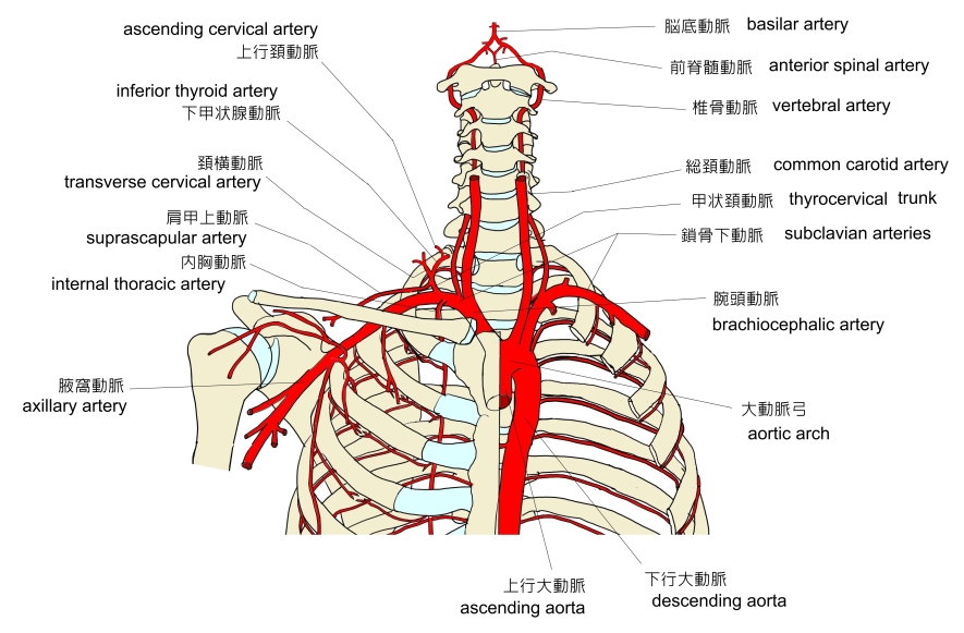
鎖骨下動脈とは

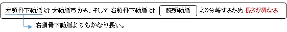
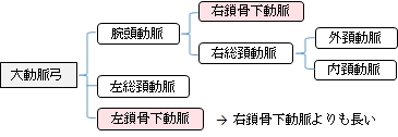

1 . 分岐後(右鎖骨下動脈は腕頭動脈、左鎖骨下動脈は動脈弓から)強膜頂の上を外側方に弓状に走る。
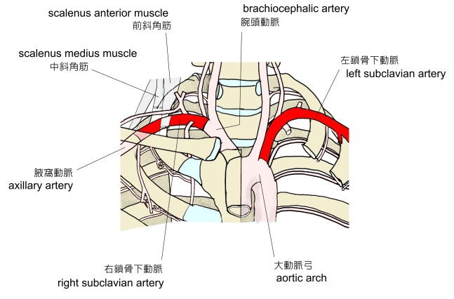
2 . 前斜角筋と中斜角筋との間(斜角筋隙)を通過する。
3 . 鎖骨下動脈溝(第1肋骨の上面)を通って鎖骨の下に達する。
4 . 鎖骨および鎖骨下筋の下(第1肋骨の外側縁)で腋窩動脈に移行する。

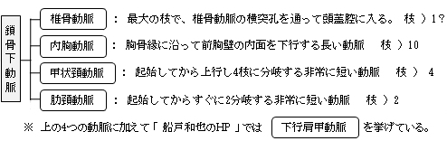
以下は鎖骨下動脈の枝を簡単に表した図となる。


以下は「 Wikipedia 」の解説文となる。
「 In human anatomy, the subclavian arteries are paired major arteriesof the upper thorax, below the clavicle. They receive blood from the aortic arch. The left subclavian artery supplies blood to the left arm and the right subclavian artery supplies blood to the right arm, with some branches supplying the head and thorax. On the left side of the body, the subclavian comes directly off the aortic arch, while on the right side it arises from the relatively short brachiocephalic artery when it bifurcates into the subclavian and the right common carotid artery.
The usual branches of the subclavian on both sides of the body are the vertebral artery, the internal thoracic artery, the thyrocervical trunk, the costocervical trunk and the dorsal scapular artery, which may branch off the transverse cervical artery which is a branch of the thyrocervical trunk. The subclavian becomes the axillary artery at the lateral border of the first rib.
【 語 句 】
・ dorsal scapular artery:肩甲背動脈 ・ transverse cervical artery :頚横動脈 ・ axillary artery :腋窩動脈)
【 structure 】
From its origin, the subclavian artery travels laterally, passing between anterior and middle scalene muscles, with the anterior scalene (scalenus anterior) on its anterior side and the middle scalene (scalenus medius) on its posterior. This is in contrast to the subclavian vein, which travels anterior to the scalenus anterior. As the subclavian artery crosses the lateral border of the first rib, it becomes the axillary artery.
On the right side the subclavian artery arises from the brachiocephalic (innominate) artery behind the right sternoclavicular articulation ; on the left side it springs from the arch of the aorta. The two vessels, therefore, in the first part of their course, differ in length, direction, and relation with neighboring structures.
In order to facilitate the description, each subclavian artery is divided into three parts:
- ・ The first portion extends from the origin of the vessel to the medial border of the Scalenus anterior.
- ・ The second lies behind this muscle.
- ・ The third extends from the lateral margin of the muscle to the outer border of the first rib, where it becomes the axillary artery.
The first portions of the two vessels require separate descriptions; the second and third parts of the two arteries are practically alike. 」
【 語 句 】
・ sternoclavicular articulation : 胸鎖関節 ・ facilitate : 容易にする ・ description : 記述、解説
【 イラスト掲載サイト 】
・ イラストや写真を掲載しているサイト-Ⅰ
・ イラストや写真を掲載しているサイト-Ⅱ
・ イラストや写真を掲載しているサイト-Ⅲ
・ イラストや写真を掲載しているサイト-Ⅳ
・ イラストや写真を掲載しているサイト-Ⅴ(枝の分岐のバリエーション)

|