・「 弁は存在しない。」( Wikipedia )


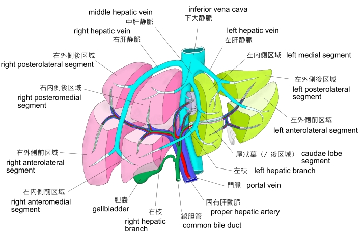
・「 通常は左肝静脈と中肝静脈は合して1本となって下大静脈にそそぐことが多い。」( 船戸和弥のHP )
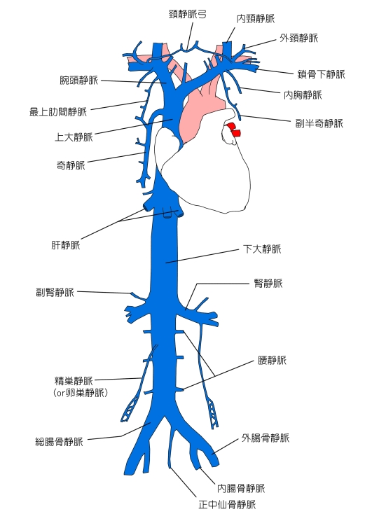
【 肝静脈が下大静脈に注ぐ位置 】
「 日本人体解剖学 」に次のような解説が見られる。
「 下大静脈が肝臓後縁の大静脈溝を通過する部位に開口する。」
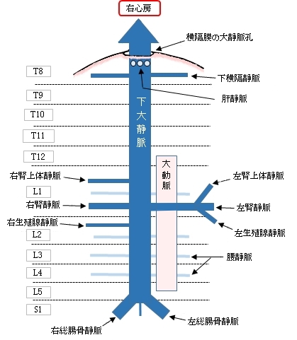
ほぼ 第8胸椎 と同じくらいの高さで、大静脈孔( 横隔膜に開く下大静脈が通過する孔 )のすぐ下あたりと考えてよいと思われる。
「 船戸和弥のHP 」には以下のような解説文が見られる。
「 尾状葉からの血液は独立して下大静脈へ、あるいはまた右または左肝静脈にそそぐ。 」
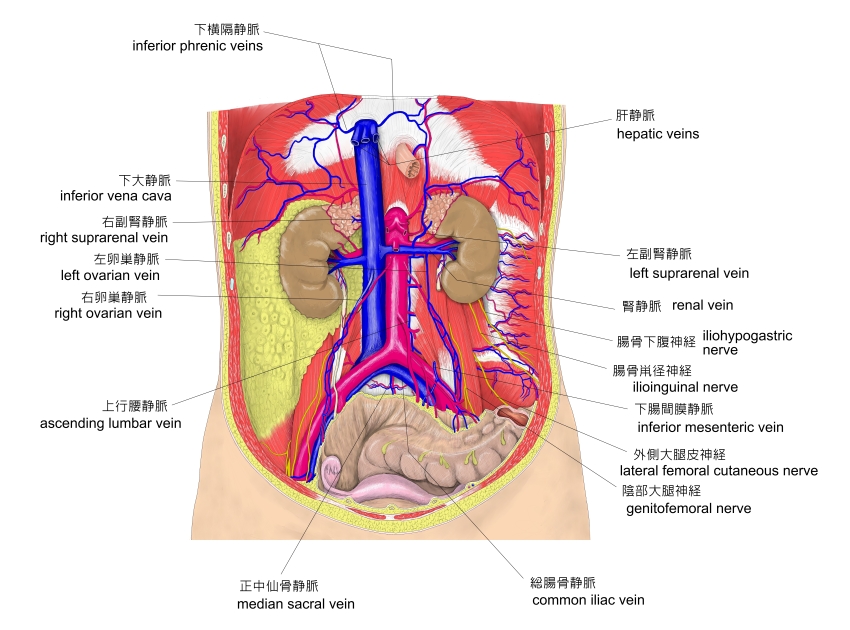
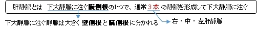
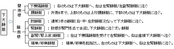
以下は下大静脈に注ぐ壁側根と臓側根の静脈を簡単に表したものになる。
以下は「 Wikipedia 」の解説文となる。
「 The hepatic veins are the veins that drain de-oxygenated blood from the liver into the inferior vena cava. There are usually three upper hepatic veins draining from the left, middle, and right parts of the liver. These are larger than the group of lower hepatic veins that can number from six to twenty. All of the hepatic veins drain into the inferior vena cava.[1]
They are one of two sets of veins connected to the liver, the others are the portal veins.
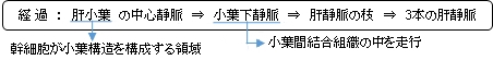
The large hepatic veins arise from smaller veins found within the liver, and ultimately from numerous central veins of the liver lobules. None of the hepatic veins have valves.
【 Structure 】
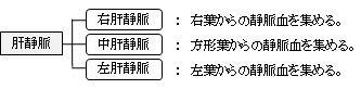
The hepatic veins are divided into an upper and a lower group. The upper three drain the central veins from the right, middle, and left regions of the liver and are larger than the lower group of veins.[1]
The lower group of from six to twenty smaller hepatic veins come from the right lobe and the caudate lobe, are in contact with the hepatic tissue, and are valveless. All the veins empty into the inferior vena cava at the back of the liver.[1] 」
【 語 句 】
・ de-oxygenated : 脱酸素化した ・ inferior vena cava : 下大静脈 ・ portal veins : 門脈 ・ caudate lobe : 尾状葉
【 イラスト掲載サイト 】
・ イラストや写真を掲載しているサイト-Ⅰ
・ イラストや写真を掲載しているサイト-Ⅱ
・ イラストや写真を掲載しているサイト-Ⅲ
・ イラストや写真を掲載しているサイト-Ⅳ
・ イラストや写真を掲載しているサイト-Ⅴ

|