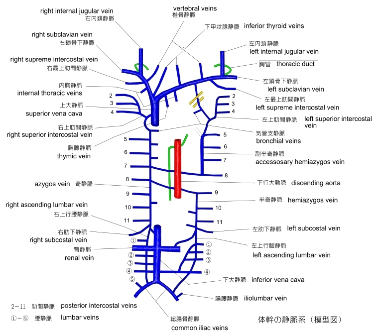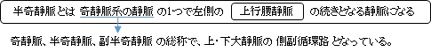

・ 半奇静脈の上方に続く部分を 副半奇静脈 ( accessory hemiazygos vein ) といい、その上端は左最上肋間静脈に続く。

【 経路詳細 】 参考 :「 船戸和弥のHP 」
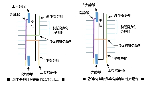
1 . 左上行腰静脈の続きとして始まる。
( 起始する高さに言及している資料は見当たらなかったが、インターネットで画像検索をしてみると、奇静脈と
同じ高さ( 第1 または 第2腰椎 )で起始しているイラストが多く見受けられた。)
2 . 上行して横隔膜 ( diaphragm ) の左脚 ( left crus ) を貫いて胸腔に入る。
3 . 脊柱の左側を上行し、第9胸椎の高さで右方向に向きを変える。
4 . 下行大動脈、食道 ( esophagus ) 、胸管 ( thoracic duct ) の後方を通って脊柱の右側に至り奇静脈に合流する。
「 日本人体解剖学 」には「 異常 」ということで以下のような解説文が見受けられる。
「 半奇静脈が、大動脈の前を通り奇静脈に向かうことがある。ときには、半奇静脈および副半奇静脈を欠き1本の奇静脈が正中線よりやや右側で胸椎の前を上り、両側の肋間静脈を受け入れることもある。」
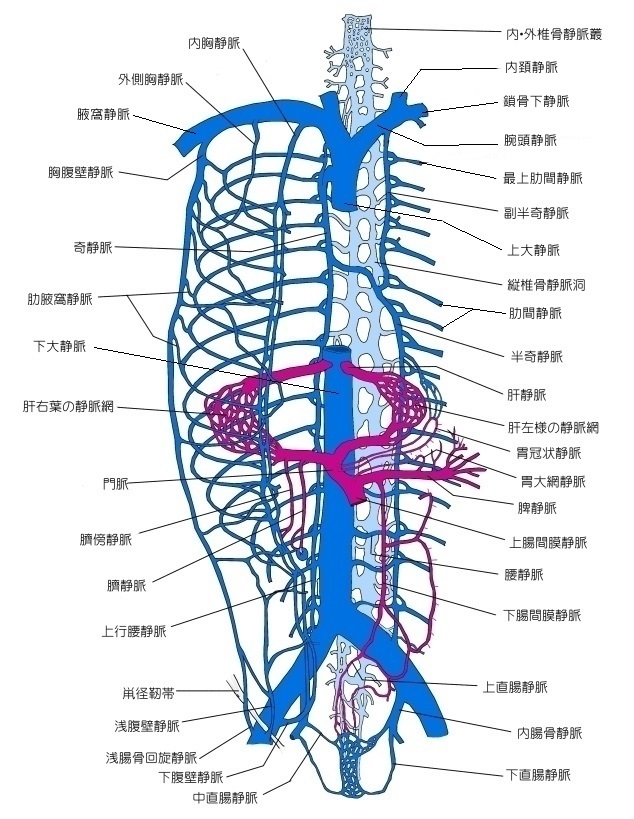
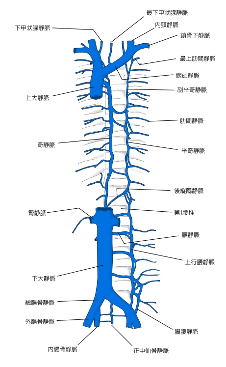
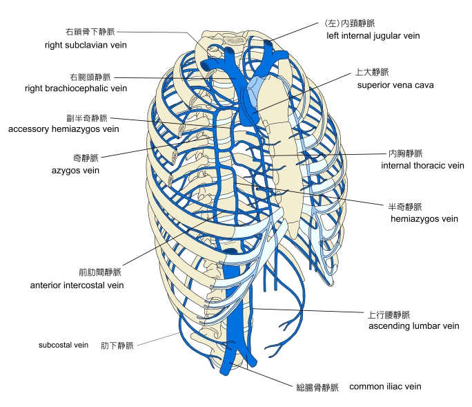
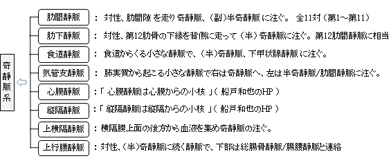
以下は奇静脈系に注ぐ静脈となる。
以下は「 Wikipedia 」の解説文となる。
「 The hemiazygos vein ( vena azygos minor inferior ) is a vein running superiorly in the lower thoracic region, just to the left side of the vertebral column.
【 Structure 】
The hemiazygos vein and the accessory hemiazygos vein, when taken together, essentially serve as the left-sided equivalent of the azygos vein. That is, the azygos vein serves to drain most of the posterior intercostal veins on the right side of the body, and the hemiazygos vein and the accessory hemiazygos vein drain most of the posterior intercostal veins on the left side of the body. Specifically, the hemiazygos vein mirrors the bottom part of the azygos vein.
The structure of the hemiazygos vein is often variable. It usually begins in the left ascending lumbar vein or renal vein, and passes upward through the left crus of the diaphragm to enter the thorax. It continues ascending on the left side of the vertebral column, and around the level of the ninth thoracic vertebra, it passes rightward across the column, behind the aorta, esophagus, and thoracic duct, to end in the azygos vein.
【 語 句 】
・ thoracic region : 胸部 ・ vertebral column : 脊柱 ・ accessory hemiazygos vein : 副半奇静脈 ・ equivalent : 同等の ・ posterior intercostal veins : 肋間静脈 ・ ascending lumbar vein : 上行腰静脈 ・ renal vein : 腎静脈 ・ left crus of the diaphragm : 横隔膜左脚 ・ thorax : 胸郭 ・ thoracic vertebra : 胸椎 ・ aorta : 大動脈 ・ esophagus : 食道 ・ thoracic duct : 胸管
The hemiazygos may or may not be continuous superiorly with the accessory hemiazygos vein.
It receives the 9th, 10th, and 11th posterior intercostal veins and the subcostal vein of the left side, and some esophageal and mediastinal veins.
The dilated hemiazygos system displayed by chest or abdominal X-ray films can be misdiagnosed as a mediastinal or retroperitoneal neoplasm, lymphadenopathy or aortic dissection (2, 5–7, 10). Venostasis which is the consequence of pathological conditions such as acquired obstruction of the IVC or SVC, the right heart failure, portal hypertension or due to pregnancy can have the same clinical presentation as hemiazygos continuation of the IVC (5, 6, 10, 13). In the case of hemiazygos continuation of the IVC, the hepatic veins can drain directly into the right atrium (3, 10). An incidental finding of this condition during venous cannulation for cardiopulmonary bypass complicates the procedure, since no solid IVC trunk for placing the cannula is present. Separate cannulation of the SVC and the right atrium should be used in this case.[2]」
【 語 句 】
・ subcostal vein : 肋下静脈 ・ esophageal vein : 食道静脈 ・ mediastinal vein : 縦隔静脈 ・ dilated : 拡張した? ・ retroperitoneal neoplasm : 後腹膜腫瘍 ・ lymphadenopathy : リンパ節腫 ・ aortic dissection : 大動脈解離 ・ Venostasis : 静脈うっ血 ・ consequence : 重要さ ・ pathological : 病理学上の ・ IVC : inferior vena cava 下大静脈 ・ SVC : superior vena cava 上大静脈 ・ portal hypertension : 門脈圧亢進 ・hepatic veins :肝静脈 ・ right atrium:右心房 ・ cannulation:カニューレ挿入 ・ cardiopulmonary bypass : 心肺バイパス術 ・ complicate : わかりにくくする
【 イラスト掲載サイト 】
・ イラストや写真を掲載しているサイト-Ⅰ
・ イラストや写真を掲載しているサイト-Ⅱ
・ イラストや写真を掲載しているサイト-Ⅲ
・ イラストや写真を掲載しているサイト-Ⅳ
・ イラストや写真を掲載しているサイト-Ⅴ
|