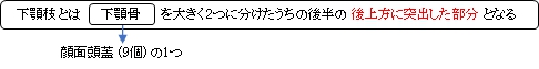
下顎枝 ( かがくし、 英:ramus of mandible )

 |
 |
 |
 |
|
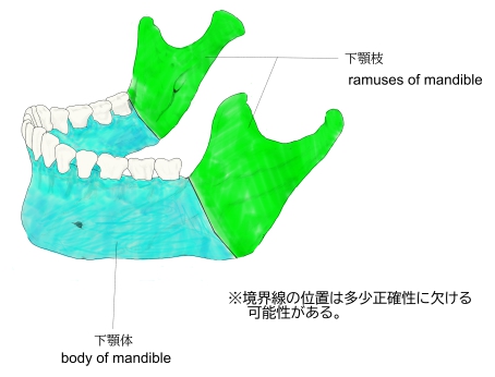
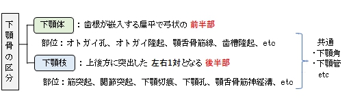
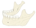
下顎体・下顎枝 |
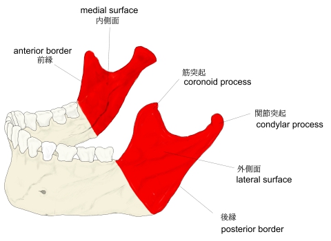
下顎枝 |
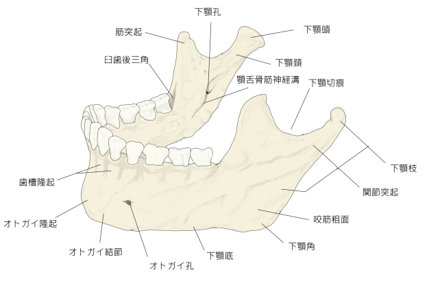
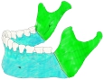
下顎骨(全体) |
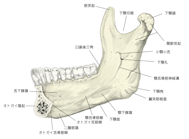
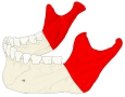
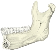
下顎骨(内側面) |
|
以下は「Wikipedia」の解説文となる。
The ramus (Latin: branch) of the human mandible has four sides, two surfaces, four borders, and two processes.
On the outside, the ramus is flat and marked by oblique ridges at its lower part. It gives attachment throughout nearly the whole of its extent to the masseter muscle.
On the inside at the center there is an oblique mandibular foramen, for the entrance of the inferior alveolar vessels and nerve. The margin of this opening is irregular; it presents in front a prominent ridge, surmounted by a sharp spine, the lingula of the mandible, which gives attachment to the sphenomandibular ligament; at its lower and back part is a notch from which the mylohyoid groove runs obliquely downward and forward, and lodges the mylohyoid vessels and nerve. Behind this groove is a rough surface, for the insertion of the medial pterygoid muscle. The mandibular canal runs obliquely downward and forward in the ramus, and then horizontally forward in the body, where it is placed under the alveoli and communicates with them by small openings. On arriving at the incisor teeth, it turns back to communicate with the mental foramen, giving off two small canals which run to the cavities containing the incisor teeth. In the posterior two-thirds of the bone the canal is situated nearer the internal surface of the mandible; and in the anterior third, nearer its external surface. It contains the inferior alveolar vessels and nerve, from which branches are distributed to the teeth.
【 語 句 】
・oblique:斜めの ・ridge:隆起線 ・masseter muscle:咬筋 ・mandibular foramen:下顎孔 ・inferior alveolar vessels:下歯槽動・静脈 ・prominent:突出した ・lingula of the mandible:下顎小舌 ・sphenomandibular ligament:蝶下顎靭帯 ・mylohyoid groove:顎舌骨筋神経溝 ・mylohyoid vessels:顎舌骨筋動・静脈 ・medial pterygoid muscle:内側翼突筋 ・mandibular canal:下顎管 ・alveoli:歯槽 ・incisor teeth:門歯
Borders
- The lower border of the ramus is thick, straight, and continuous with the inferior border of the body of the bone. At its junction with the posterior border is the angle of the mandible, which may be either inverted or everted and is marked by rough, oblique ridges on each side, for the attachment of the masseter laterally, and the medial pterygoid muscle medially; the stylomandibular ligament is attached to the angle between these muscles. The anterior border is thin above, thicker below, and continuous with the oblique line.
- The region where the lower border meets the posterior border is the angle of the mandible, often called the gonial angle.
- The posterior border is thick, smooth, rounded, and covered by the parotid gland. The upper border is thin, and is surmounted by two processes, the coronoid in front and the condyloid behind, separated by a deep concavity, the mandibular notch.
Processes
- The coronoid process is a thin, triangular eminence, which is flattened from side to side and varies in shape and size.
- The condyloid process is thicker than the coronoid, and consists of two portions: the mandibular condyle, and the constricted portion which supports it, the neck. The condyle is the most superior part of the mandible and is part of the temporomandibular joint.
- The mandibular notch, separating the two processes, is a deep semilunar depression and is crossed by the masseteric vessels and nerve.
【 語 句 】
・angle of the mandible:下顎角 ・stylomandibular ligament:茎突下顎靭帯 ・oblique line:斜線 ・gonial angle:下顎角 ・parotid gland:耳下腺 ・surmount:載せる ・coronoid process:筋突起 ・condyloid process:関節突起 ・concavity:くぼみ ・mandibular notch:下顎切痕 ・constricted:圧縮させられた? ・temporomandibular joint:顎関節 ・semilunar:三日月形の
■ 写真やイラストを掲載しているサイト ■
・ イラストや写真を掲載しているサイト-Ⅰ
・ イラストや写真を掲載しているサイト-Ⅱ
・ イラストや写真を掲載しているサイト-Ⅲ
・ イラストや写真を掲載しているサイト-Ⅳ
|
|