

外腹斜筋( がいふくしゃきん、英:External oblique muscle )
・ 概 要 |
・ 作 用 |
・ イラスト掲載サイト |
|
・ イラスト |
・ 神経 / 脈管 |
||
・ 起始 / 停止 |
・ Wikipedia |
![]()

![]()
・「 胸部における 「外肋間筋」 に相当する。」 ( 日本人体解剖学 )
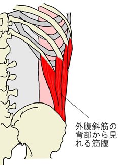
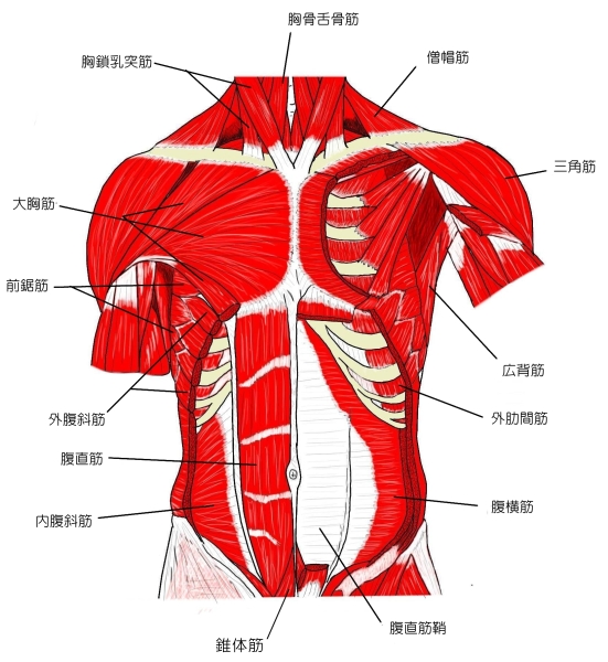
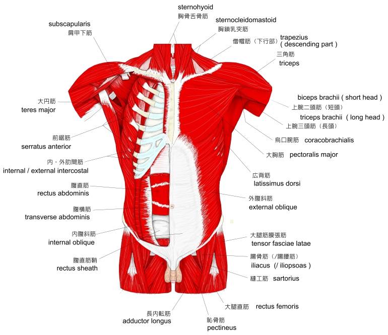
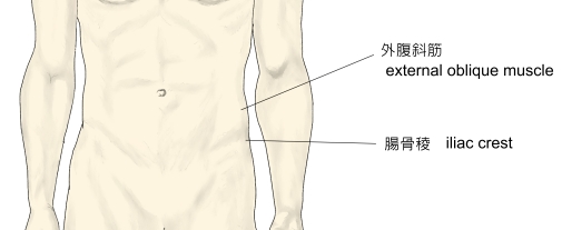
・ 筋が体幹の外側面を斜めに走っていて、その筋腹は側腹部の前下部と鼡径部には見られない。⇒ イラスト
・ 一部は、体表部において前鋸筋と楔型に交わっている。 ⇒ イラスト ( 日本人体解剖学 )
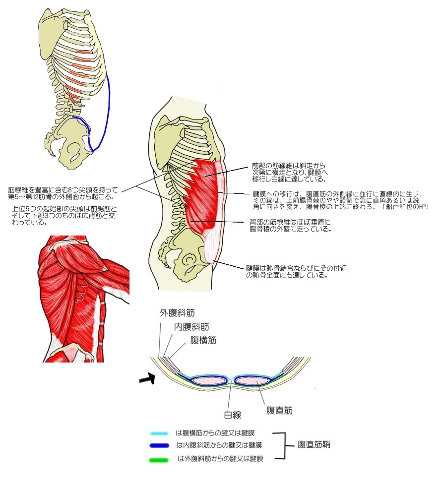
以下は 「船戸和也のHP」 の解説文の一部となる。
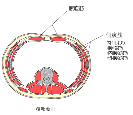
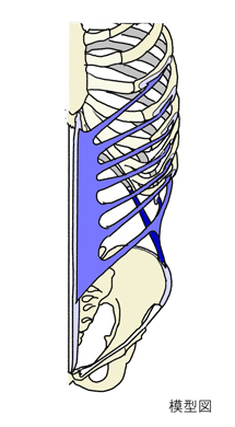
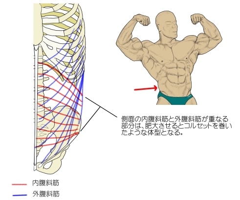
「 3つの側腹筋の中でもっとも広い領域を占める筋である。最初の起始筋束は、わずかに斜めに下る程度であるが、もっとも尾側の筋束は急角度で下方へ進む。
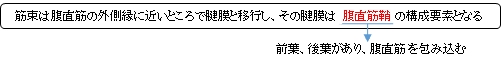
腱膜への移行は、腹直筋の外側縁に並行に直線的に生じ、その線は、上前腸骨棘のやや頭側で急に直角あるいは鋭角に向きを変え、腸骨稜の上端に終わる。このようにして筋角が形成されるが、この角はしばしば皮膚の表面から認められ、古代の彫刻にも良く強調されている。この筋線維束は、筋が引き伸ばされない限りは腸骨稜を越えて膨出し、そのために骨盤の上縁をおおって鼡径隆起を形成する。多くの人において(男性より女性において頻度が高いが)広背筋は外腹斜筋に直接隣接しない。広背筋の前縁と外腹斜筋の後縁の間には尾側が腸骨稜によって境される三角形の領域、腰三角が形成され、この部位の腹壁は、内服斜筋と腹横筋にのみによって形成される。ここは、脊柱に生じ腸骨稜に沿って移動してきた膿瘍が、外へ逃げることが出きる場所であり、まれには(下)腰ヘルニアも出現する。」
また、「Rauber-Kopsch解剖学」 では以下のような解説が見られる。
「 変 異 : (M. obliquus abdominis externus profundusの1例が報告されている(中山知雄,実田茂=解剖学雑誌,27巻,89~94,1952)完全欠如はまだみられていないが,退化的になっていることはある.筋が重複していることはまれである.起始尖頭の数は7個のことがあり,また9個に達することがある;その最下の起始は腰背筋膜および第1腰椎の肋骨突起から起っている.存在の不定な深部の起始尖頭が上部の弓肋の前端からでている.」
![]()
 |
 |
 |
 |
 |
 |
 |
 |
 |
![]()
【 起 始 】 : 第5~第12肋骨の外側面
起始後、筋束はほぼ並行するように斜め下前方に走る。
【 停 止 】 : ・腸骨の腸骨稜の外唇の前半、 鼡径靭帯、 白線
 |
 |
|
|
![]()
・体幹部の前屈及び同側方向に側屈。 ・胸郭を引き下げる。
・骨盤の前縁部を引き上げる。
![]()
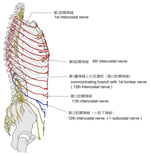
・神 経:肋間神経 (Th5~Th12)
※ 資料によっては(T7~T12)または、左記にプラスして腸骨下腹神経、腸骨鼡径神経が加わる
ことがある。
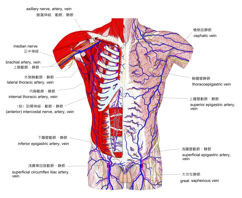
胸部~腹部の血管 |
![]()
The abdominal external oblique muscle (also external oblique muscle, or exterior oblique) is the largest and outermost of the three flat abdominal muscles of the lateral anterior abdomen.
【 Structure 】
The external oblique is situated on the lateral and anterior parts of the abdomen. It is broad, thin, and irregularly quadrilateral, its muscular portion occupying the side, its aponeurosis the anterior wall of the abdomen. In most humans (especially females), the oblique is not visible, due to subcutaneous fat deposits and the small size of the muscle.
It arises from eight fleshy digitations, each from the external surfaces and inferior borders of the fifth to twelfth ribs (lower eight ribs). These digitations are arranged in an oblique line which runs inferiorly and anteriorly, with the upper digitations being attached close to the cartilages of the corresponding ribs, the lowest to the apex of the cartilage of the last rib, the intermediate ones to the ribs at some distance from their cartilages.
The five superior serrations increase in size from above downward, and are received between corresponding processes of the serratus anterior muscle; the three lower ones diminish in size from above downward and receive between them corresponding processes from the latissimus dorsi. From these attachments the fleshy fibers proceed in various directions. Its posterior fibers from the ribs to the iliac crest form a free posterior border.
Those from the lowest ribs pass nearly vertically downward, and are inserted into the anterior half of the outer lip of the iliac crest; the middle and upper fibers, directed downward (inferiorly) and forward (anteriorly), become aponeurotic at approximately the midclavicular line and form the anterior layer of the rectus sheath. This aponeurosis formed from fibres from either side of the external oblique decussates at the linea alba.
The aponeurosis of the external oblique muscle forms the inguinal ligament. The muscle also contributes to the inguinal canal.
The internal oblique muscle is just deep to the external oblique muscle.
【 Nerve supply 】
The external oblique muscle is supplied by ventral branches of the lower six thoracoabdominal nerves and the subcostal nerve on each side.
【 Blood supply 】
The cranial portion of the muscle is supplied by the lower intercostal arteries, whereas the caudal portion is supplied by a branches of either the deep circumflex iliac artery or the iliolumbar artery.
【 Function 】
The external oblique functions to pull the chest downwards and compress the abdominal cavity, which increases the intra-abdominal pressure as in a valsalva maneuver. It also performs ipsilateral (same side) side-bending and contralateral (opposite side) rotation. So the right external oblique would side bend to the right and rotate to the left. The internal oblique muscle functions similarly except it rotates ipsilaterally.
【 語 句 】
・ abdominal muscles : 腹筋 ・ abdomen : 腹部 ・ quadrilateral : 四辺形の ・ aponeurosis : 腱膜 ・ subcutaneous : 皮下の ・ fleshy : 肉質の ・ digitation : 指状突起 ・ serration : 鋸歯状 ・ serratus anterior muscle : 前鋸筋 ・ diminish : 減らす ・ latissimus dorsi : 広背筋 ・ iliac crest : 腸骨稜 ・ approximately : おおよそ ・ midclavicular line : 鎖骨中線 ・ rectus sheath : 腹直筋鞘 ・ decussates : X字形の ・linea alba : 白線 ・ inguinal ligament : 鼡径靭帯 ・ contribute : 寄与する ・ inguinal canal : 鼡径管 ・ internal oblique muscle : 内腹斜筋 ・ ventral branches : 腹側枝 ・ thoracoabdominal nerves : 胸腹神経? ・ subcostal nerve : 肋下神経 ・ cranial : 頭蓋の ・ intercostal arteries : 肋間動脈 ・ caudal : 尾部の ・ deep circumflex iliac artery : 深腸骨回旋動脈 ・ iliolumbar artery : 腸腰動脈 ・ compress : 圧縮する ・ abdominal cavity : 腹腔 ・ valsalva maneuver : バルサルバ法 ・ ipsilateral : 同側の ・contralateral : 対側の
![]()
![]()