

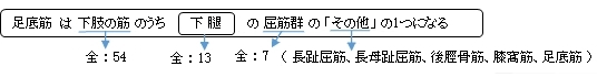
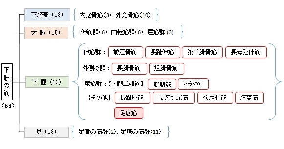
足底筋 ( そくていきん、英: plantaris muscle )
・ 概 要 |
・ 作 用 |
・ イラスト掲載サイ |
|
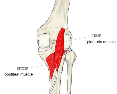
・ イラスト |
・ 神経 / 脈管 |
||
・ 起始 / 停止 |
・ Wikipedia |
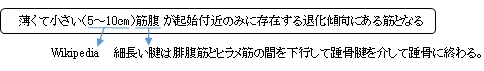
・Wikipediaでは腱の長さを「人体の中で最長で、その長さは30~45㎝」としている。
以下は「船戸和也のHP」から抜粋した解説文となる。
・腓腹筋とヒラメ筋の間にあり、結合組織に囲まれている筋。
・ときどき下腿筋膜に、あるいはまれには足底腱膜に続く。
・足底腱膜へ続く筋の、系統発生的な、遺残筋である。
・ときどき欠如したり(8~12%「Wikipedia」)腓腹筋外側頭と癒合したりする。
以下は「船戸和也のHP」の解説文となる。
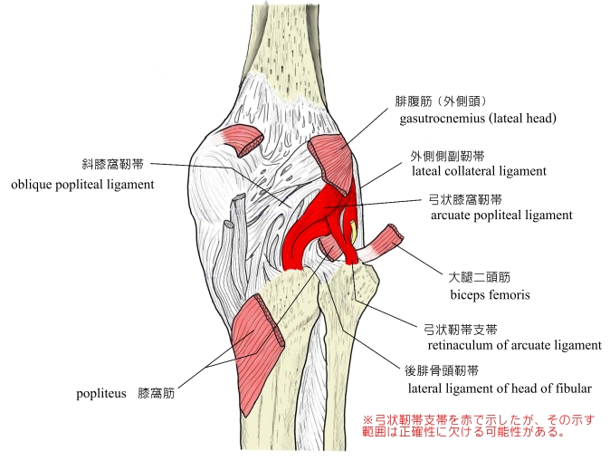
「足底筋は腓腹筋とヒラメ筋の間にあり、結合組織に囲まれている筋。腓腹筋外側頭の内側、大腿骨外側顆の外側上顆稜から起こる。細い筋腹から細く長い停止腱が腓腹筋とヒラメ筋の間を走り、(アキレス腱を介して)踵骨隆起へ至る。同筋はときどき下腿筋膜に、あるいはまれには足底腱膜に続く。足底筋は下等霊長類にみられる足底腱膜へ続く筋の、系統発生的な、遺残筋である。人ではときどき欠如したり腓腹筋外側頭と癒合したりする。足底筋は腓腹筋とヒラメ筋の間にあって、結合組織に包まれている。その結合組織は後脛骨動静脈の血管鞘と連絡している。同筋はヒラメ筋腱弓よりも上ではこれらの血管を守るようにその上を走る。この筋は脛骨神経支配を受け、距腿関節による足の底屈、膝関節の屈曲を助けるが、その力は弱い。膝関節屈曲中に足底筋は神経血管索を引き上げ、弓状走行をとらせ、血管が折れ曲がるのを防ぐ役割をする。) 」
 |
|
 |
【 起 始 】 : 外側上顆(大腿骨)、膝関節包(特に弓状膝窩靭帯から)
【 停 止 】 : 踵骨腱(アキレス腱)を介し踵骨隆起(踵骨)に至る。
※「下腿筋膜、足底筋膜にも停止する」(日本人体解剖学)
※「ときおり独立した腱が踵骨に停止する。」(Wikipedia)
「下腿三頭筋の作用を助ける。 」 (日本人体解剖学)
※退化傾向にある筋のため、この筋単独での働きはそれほど強くない。
・ 神 経 : 脛骨神経(L4,L5,S1)
The plantaris is one of the superficial muscles of the superficial posterior compartment of the leg, one of the fascial compartments of the leg.
It is composed of a thin muscle belly and a long thin tendon. While not as thick as the achilles tendon, the plantaris tendon (which tends to be between 30–45 centimetres (12–18 in) in length) is the longest tendon in the human body. Not including the tendon, the plantaris muscle is approximately 5–10 centimetres (2.0–3.9 in) long and is absent in 8-12% of the population. It is one of the plantar flexors in the posterior compartment of the leg, along with the gastrocnemius and soleus muscles. The plantaris is considered an unimportant muscle and mainly acts with the gastrocnemius.
【Structure】
The plantaris muscle arises from the inferior part of the lateral supracondylar ridge of the femur at a position slightly superior to the origin of the lateral head of gastrocnemius. It passes posterior to the knee joint in an inferomedial direction and becomes tendinous distally to insert into the Achilles tendon. It occasionally separately inserts into the medial side of the calcaneus.
【Innervation】
The plantaris muscle is innervated by the tibial nerve, a branch of the sciatic nerve in the sacral plexus. Signaling for contraction begins in the frontal lobe of the brain with the pre-central gyrus (primary motor cortex). Upper motor neurons are stimulated and send a signal through the internal capsule and down the corticospinal tract. Decussation of the lateral corticospinal tract occurs in the medullary pyramids, then the fibers continue down the contralateral side of the spinal cord. Upper motor neurons synapse with lower motor neurons at the anterior horn of the spinal cord in the sacral plexus (formed from the anterior rami of spinal nerves L4, L5, S1–4). The lower motor neuron fibers continue down the sciatic nerve and then diverge into the tibial and common fibular nerves. The tibial nerve runs medially at the knee joint. When the tibial nerve receives an action potential, the plantaris muscle contracts, providing weak plantar flexion of the foot and weak flexion of the knee.
【Variation】
The muscle may arise from the oblique popliteal ligament. Interdigitations with the lateral head of the gastrocnemius and a fibrous extension of the muscle to the patella are not unusual.
【Function】
The plantaris acts to weakly plantar flex the ankle joint and flex the knee joint.
The plantaris muscle may also provide proprioceptive feedback information to the central nervous system regarding the position of the foot. The unusually high density of proprioceptive receptor end organs supports this notion.
Its motor function is so minimal that its long tendon can readily be harvested for reconstruction elsewhere with little functional deficit. Often mistaken for a nerve by new medical students (and thus called the "freshman's nerve"), the muscle was useful to other primates for grasping with their feet.
【 語 句 】
・achilles tendon:アキレス腱 ・gastrocnemius:腓腹筋 ・soleus:ヒラメ筋 ・lateral supracondylar ridge:外側上顆稜 ・femur:大腿骨 ・calcaneus:踵骨 ・tibial nerve:脛骨 ・sciatic nerve:坐骨神経 ・sacral plexus:仙骨神経叢 ・frontal lobe:前頭葉 ・pre-central gyrus:中心前回 ・primary motor cortex:一次運動野 ・corticospinal tract:皮質脊髄路 ・decussation:交差 ・medullary pyramid:腎錐体 ・contralateral:反対側の ・motor neurons:運動神経細胞 ・anterior horn:前角 ・diverge:分岐する、広がる ・contract:収縮する ・oblique popliteal ligament: ・Interdigitation:嵌合 ・patella:膝蓋骨 ・proprioceptive:固有受容性の ・high density:高密度 ・deficit:不足 ・primate:霊長類
![]()