

大胸筋 ( だいきょうきん、英:pectoralis major muscle )
・ 概 要 |
・ 作 用 |
・ イラスト掲載サイト |
|
・ イラスト |
・ 神経 / 脈管 |
||
・ 起始 / 停止 |
・ Wikipedia |
![]()
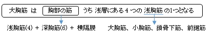
・ 「 起始部の表面に稀に 胸骨筋 という筋が見られることがある。( 日本人の約10% ) 」 (参考:日本人体解剖学)
![]()
【 関連語句 】
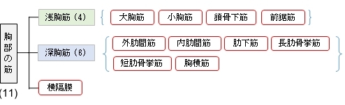
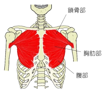
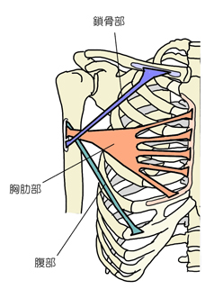
大胸筋は以下の3つの溝および空隙の形成にあずかる。
・ 胸筋間溝 : 鎖骨部と胸肋部の間の溝
・ 三角筋大胸筋溝 : 三角筋胸筋三角下部の三角筋と大胸筋の鎖骨部との境界にあたる溝
・ 三角筋大胸筋三角 : 鎖骨下部の鎖骨、三角筋、および大胸筋の鎖骨部で囲まれた三角状の窪み
・ 「 船戸和弥のHP 」 の 「 Rauber-Kopsch解剖学 」 ではこの筋の解説において「変異」ということで解説が見られる。
⇒ 解説のページへ
筋連結 : 腹直筋、 三角筋、 胸鎖乳突筋 ・反対側の大胸筋
![]()
 |
 |
|
 |
 |
![]()
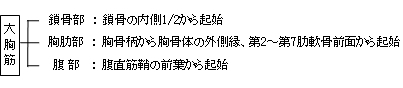
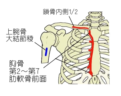
【 起 始 】 : ・ 鎖骨部 : 鎖骨の内側1/2
・ 胸肋部 : 胸骨柄から胸骨体の外側縁、第2~第7肋軟骨前面
・ 腹 部 : 腹直筋鞘の前葉
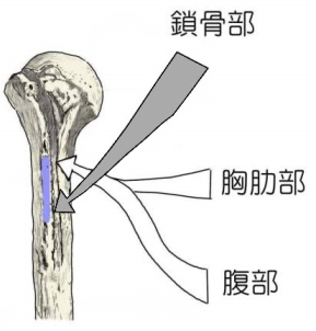
【 停 止 】 : 上腕骨、大結節稜 ( 停止部の幅は約5㎝ 【 Wikipedia 】 )
※ 停止部では鎖骨部はその遠位に終わり、胸肋部と腹部では鎖骨部の下を交差する形で近位に終わる。 ⇒ イラスト
 |
 |
 |
![]()
上腕の内転及び内旋 ・上腕を固定した時、胸骨と肋骨を引き上げる。
![]()
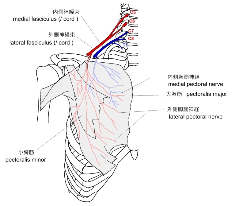
・ 神 経 : 内側胸筋神経 (C7, C8)、 外側胸筋神経 (C5~C8, T1)
・ 脈 管 :
 |
![]()
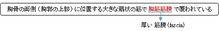
The pectoralis major (from Latin pectus 'breast') is a thick, fan-shaped muscle, situated at the chest of the human body. It makes up the bulk of the chest muscles and lies under the breast. Beneath the pectoralis major is the pectoralis minor, a thin, triangular muscle. The pectoralis major's primary functions are flexion, adduction, and internal rotation of the humerus. The pectoral major may colloquially be referred to as "pecs", "pectoral muscle" or "chest muscle" due to it being the largest and most superficial muscle in the chest area.
【 Structure 】
It arises from the anterior surface of the sternal half of the clavicle from breadth of the half of the anterior surface of the sternum, as low down as the attachment of the cartilage of the sixth or seventh rib; from the cartilages of all the true ribs, with the exception, frequently, of the first or seventh, and from the aponeurosis of the abdominal external oblique muscle.
From this extensive origin the fibers converge toward their insertion; those arising from the clavicle pass obliquely downward and outwards (laterally), and are usually separated from the rest by a slight interval; those from the lower part of the sternum, and the cartilages of the lower true ribs, run upward and laterally, while the middle fibers pass horizontally.
They all end in a flat tendon, about 5 cm in breadth, which is inserted into the lateral lip of the bicipital groove (intertubercular sulcus) of the humerus.[clarification needed]
This tendon consists of two laminae, placed one in front of the other, and usually blended together below:
- The anterior lamina, which is thicker, receives the clavicular and the uppermost sternal fibers. They are inserted in the same order as that in which they arise: the most lateral of the clavicular fibers are inserted at the upper part of the anterior lamina; the uppermost sternal fibers pass down to the lower part of the lamina which extends as low as the tendon of the Deltoid and joins with it.
- The posterior lamina of the tendon receives the attachment of the greater part of the sternal portion and the deep fibers, i. e., those from the costal cartilages.
These deep fibers, and particularly those from the lower costal cartilages, ascend the humerus insertion higher, turning backward successively behind the superficial and upper ones, so that the tendon appears to be twisted. The posterior lamina reaches higher on the humerus than the anterior one, and from it an expansion is given off which covers the intertubercular groove of the humerus and blends with the capsule of the shoulder-joint.
From the deepest fibers of this lamina at its insertion an expansion is given off which lines the intertubercular groove, while from the lower border of the tendon a third expansion passes downward to the fascia of the arm.
【 語 句 】
・ bulk : 大部分、大半 ・ breast : 胸 ・ flexion : 屈曲 ・ adduction : 内転 ・ internal rotation : 内旋? ・ humerus : 上腕骨 ・ colloquially : 口語で ・ clavicle : 鎖骨 ・ sternum : 胸骨 ・ cartilage : 軟骨 ・ true rib : 真肋 ・ with the exception of ~ : ~を除いて ・ aponeurosis 腱膜 ・ abdominal external oblique muscle : 外腹斜筋 ・ extensive : 広範囲に渡る ・ converge : 集中する ・ intertubercular sulcus :結節間溝 ・ laminae : 薄膜 ・ deltoid : 三角筋 ・ fascia : 筋膜
【 Nerve supply 】
The pectoralis major receives dual motor innervation by the medial pectoral nerve and the lateral pectoral nerve, also known as the lateral anterior thoracic nerve. The sternal head receives innervation from the C7, C8 and T1 nerve roots, via the lower trunk of the brachial plexus and the medial pectoral nerve. The clavicular head receives innervation from the C5 and C6 nerve roots via the upper trunk and lateral cord of the brachial plexus, which gives off the lateral pectoral nerve. The lateral pectoral nerve is distributed over the deep surface of the pectoralis major.
The sensory feedback from the pectoralis major follows the reverse path, returning via first-order neurons to the spinal nerves at C5, C6, C8, and T1 through the posterior rami. After the synapse in the posterior horn of the spinal cord, sensory information concerning movement of the muscle, proprioception, and pressure then travels through a second-order neuron in the dorsal column medial lemniscus tract to the medulla. There, the fibers decussate to form the medial lemniscus which carries the sensory information the rest of the way to the thalamus, the "gateway to the cortex". The thalamus diverts some sensory information to the cerebellum and the basal nuclei to complete the motor feedback loop while some sensory information ascends directly to the postcentral gyrus of the parietal lobe of the brain via third-order neurons. Sensory information for the pectoralis major is processed in the superior portion of the sensory homunculus, adjacent to the longitudinal fissure which divides the two hemispheres of the brain.
Electromyography suggests that it consists of at least six groups of muscle fibres that can be independently coordinated by the central nervous system.
【 Variation 】
The more frequent variations include greater or less extent of attachment to the ribs and sternum, varying size of the abdominal part or its absence, greater or less extent of separation of sternocostal and clavicular parts, fusion of clavicular part with deltoid, and decussation in front of the sternum. Deficiency or absence of the sternocostal part is not uncommon and more frequent than absence of the clavicular part.[citation needed] Poland syndrome is a rare congenital condition in which the whole muscle is missing, most commonly on one side of the body. This may accompany absence of the breast in females. The sternalis muscle may be a variant form of the pectoralis major or the rectus abdominis. [Submuscular and intramuscular surgical implants (similar to breast augmentation implants) may be available from plastic surgeons to modify aesthetic contours, mass, and asymmetry or variation in both males and females.]
【 語 句 】
・ medial pectoral nerve : 内側胸筋神経 ・ brachial plexus : 腕神経叢 ・ posterior rami : 後枝 ・ posterior horn : 後角 ・ proprioception : 固有受容性感覚 ・ medulla : 延髄 ・ decussate : X字型の ・ medial lemniscus : 内側毛体 ・ thalamus : 視床 ・ cortex : 大脳皮質 ・ cerebellum : 小脳 ・ basal nuclei : 基底核 ・ postcentral gyrus : 中心後回 ・ parietal lobe : 頭頂葉 ・ process : 処理する ・ hemisphere : 半球 ・Electromyography : 筋電図検査 ・ fusion : 連合 ・ deficiency : 欠乏 ・ congenital : 先天的な ・ sternalis muscle : 胸骨筋 ・rectus abdominis : 腹直筋 ・ augmentation : 増大 ・ modify : 修正する、緩和する ・ modify : 審美的な ・ contour : 外形 ・ mass :量 ・ asymmetry : 不均整
【 Function 】
The pectoralis major has four actions which are primarily responsible for movement of the shoulder joint. The first action is flexion of the humerus, as in throwing a ball underhand, and in lifting a child. Secondly, it adducts the humerus, as when flapping the arms. Thirdly, it rotates the humerus medially, as occurs when arm-wrestling. Fourthly the pectoralis major is also responsible for keeping the arm attached to the trunk of the body. It has two different parts which are responsible for different actions. The clavicular part is close to the deltoid muscle and contributes to flexion, horizontal adduction, and inward rotation of the humerus. When at an approximately 110 degree angle,[citation needed] it contributes to adduction of the humerus. The sternocostal part is antagonistic to the clavicular part contributing to downward and forward movement of the arm and inward rotation when accompanied by adduction. The sternal fibers can also contribute to extension, but not beyond anatomical position.
Hypertrophy of the pectoralis major increases functionality. Maximal activation of the pectoralis major occurs in the transverse plane through pressing motions. Both multi-joint and single-joint exercises induce pectoralis major hypertrophy. A combination of both single-joint and multi-joint exercises will result in a maximum hypertrophic response. [Aesthetic contours of regions in the muscle may be specifically-addressed (“targeted”) by specific exercises; for instance, “plating” or “stitching” of the pectoralis major —towards the center of the sternum —-may be targeted by a wider hand position.] The pectoralis major can be targeted from numerous training angles along the sternum and clavicle. Exercises that include horizontal adduction and elbow extensions such as the barbell bench press, dumbbell bench press, and machine bench press induce high activation of the pectoralis major in the sternocostal region. Heavy loads are strongly correlated with pectoralis major activation.
【 語 句 】
・ flexion : 屈曲 ・ adduct : 内転させる ・ antagonistic : 反対の、対立する ・ hypertrophy : 肥大 ・ induce : 誘発する ・ correlate : 互いに関係がある
![]()
![]()