

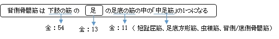
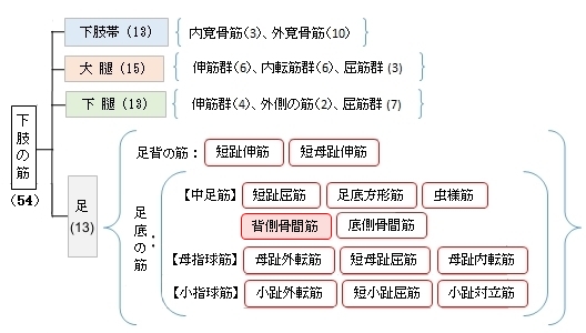
背側骨間筋 ( はいそくこつかんきん、英:dorsal interossei muscles )
・ 概 要 |
・ 作 用 |
・ イラスト掲載サイ |
|
・ イラスト |
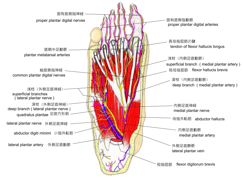
・ 神経 / 脈管 |
||
・ 起始 / 停止 |
・ Wikipedia |

 |
 |
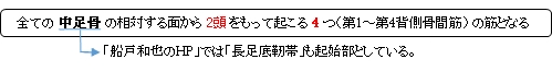
以下は「船戸和也のHP」の解説文となる。
「4つの背側骨間筋は2頭をもってすべての中足骨対向面および長足底靱帯から起こる。背側骨間筋も基節骨底に停止し、第2趾がこれらの筋の対称線となる。腱線維が指背腱膜へ放散することはほとんどない。背側骨間筋の停止腱の一部は第2~4趾の趾背腱膜展開部にも伸びる。外側足底神経の趾背を受けるこれらの筋の収縮により、第2趾を中心として散開するような各趾の外転、第2~4趾における中足趾節関節の屈曲と趾節間関節の伸展が得られる。個々の背側骨間筋が隣接する2本の中足骨より起始する関係上、この筋の収縮は中足骨を寄せ集めて前足部の構造の安定性を高める上にも役立つ。」
 |
 |
 |
 |
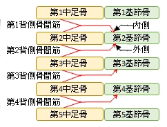
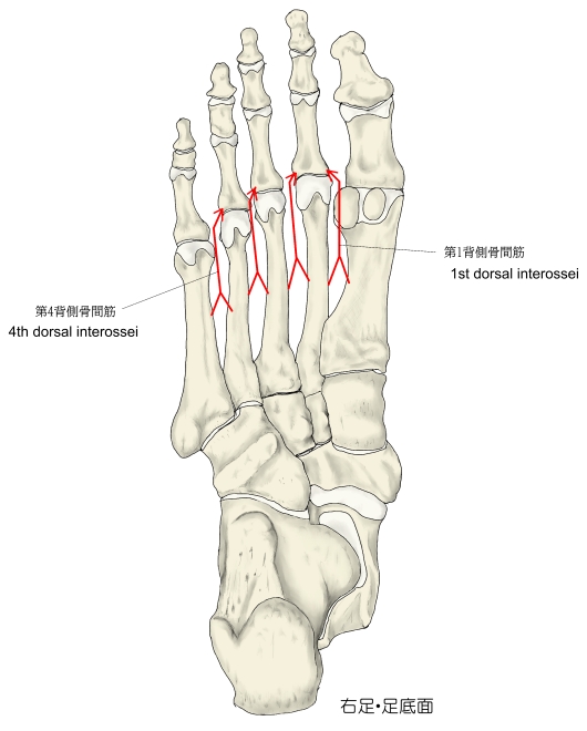
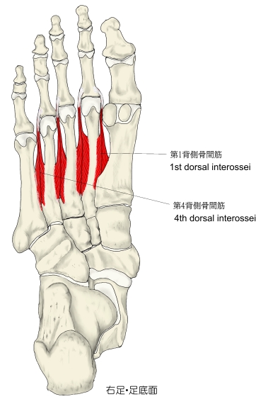
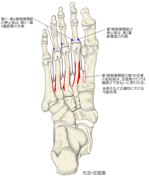
【 起 始 】: 第1~第4背側骨間筋のそれぞれが2頭を持って、第1~第5中足骨の相対する面から起こる。
【 停 止 】:・第1背側骨間筋 : 第2趾の基節骨の骨底の内側
・第2~第4背側骨間筋 : 第2~第4趾の基節骨の骨底の外側
※また、「プロメテウス解剖学アトラス 」では、基節骨の骨底以外に「指背腱膜」という記述がある。
 |
「 第1背側骨間筋は第2趾を内側方に引き、第2~第4背側骨間筋は第2~第4趾を外側方に引く。」 ( 日本人体解剖学 )
・第2~第4趾の中足趾節(MTP)関節:底屈
・第2~第4趾の近位趾節間(PIP)関節、遠位趾節間(DIP)関節:背屈
・趾を広げる。(第3、第4趾を第2趾から外転させる)
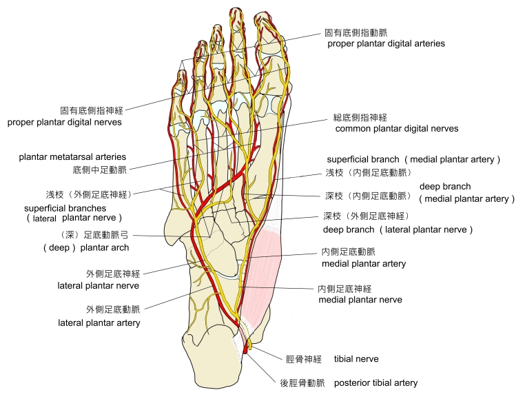
・ 神 経 : 外側足底神経(S1,S2) ※「プロメテウス解剖学アトラス 」では(S2,S3)
・ 脈 管 :
 |
 |
In human anatomy, the dorsal interossei of the foot are four muscles situated between the metatarsal bones.
【Origin】
The four interossei muscles are bipenniform muscles each originating by two heads from the proximal half of the sides of adjacent metatarsal bones.
【Insertion】
The two heads of each muscle form a central tendon which passes forwards deep to the deep transverse metatarsal ligament.The tendons are inserted on the bases of the second, third, and fourth proximal phalanges and into the aponeurosis of the tendons of the extensor digitorum longus without attaching to the extensor hoods of the toes.
Thus, the first is inserted into the medial side of the second toe; the other three are inserted into the lateral sides of the second, third, and fourth toes.
【Action】
The dorsal interossei abduct at the metatarsophalangeal joints of the third and fourth toes. Because there is a pair of dorsal interossei muscles attached on both sides of the second toe, simultaneous contraction of these muscles results in no movement. This arrangement of dorsal interossei makes the second toe the midline of the foot, whereas the midline of the hand (marked by dorsal interossei of hand) is in the third finger.
Abduction is of little importance in the foot, but, together with the plantar interossei, the dorsal interossei also produce flexion at the metatarsophalangeal joints. Although small, the dorsal interossei are powerful muscles that, together with their plantar counterparts, control the direction of the toes during violent activity, thus allowing the long and short flexors to perform their actions.
Because of the relationship to the metatarsophalangeal joints, the interossei muscles also contribute to maintaining the anterior metatarsal arch of the foot and also, to a limited extent, the medial and lateral longitudinal arches of the foot.
【Innervation】
All dorsal interossei are innervated by the lateral plantar nerve (S2–3). Those in the fourth interosseous space are innervated by the superficial branch and the other by the deep branch. The first and second dorsal interossei muscles additionally receive innervation from the lateral branch of the deep fibular nerve.
【Relations】
In the angular interval left between the heads of each of the three lateral muscles, one of the perforating arteries passes to the dorsum of the foot; through the space between the heads of the first muscle the deep plantar branch of the dorsalis pedis artery enters the sole of the foot. [3]
【 語 句 】
・metatarsal bones:中足骨 ・bipenniform:双翼状の ・proximal:近位の ・adjacent:隣接する ・transverse metatarsal ligament:深横中足靭帯 ・proximal phalanges:基節骨 ・ aponeurosis:腱膜 ・ extensor digitorum longus:長趾伸筋 ・abduct:外転させる ・metatarsophalangeal joints:中足趾節関節 ・simultaneous:同時に起こる ・contraction:収縮 ・plantar interossei:底側骨間筋 ・ lateral plantar nerve:外側足底神経 ・deep fibular nerve:深腓骨神経 ・angular:角のある ・perforating arteries:貫通動脈 ・ dorsalis pedis artery:足背動脈
![]()