

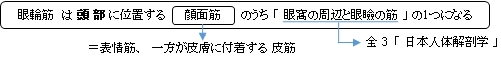
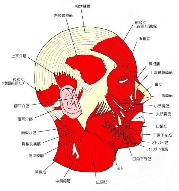
眼輪筋 ( がんりんきん、 英 : orbicularis oculi muscle ) (muscles)
・ 概 要 |
・ 作 用 |
||
・ イラスト |
・ 神経 / 脈管 |
||
・ 起始 / 停止 |
・ Wikipedia |
![]()

・ 「(異常)眼輪筋の眼窩部の一部が、深く眼窩内に入って 眼窩横筋 となることがある。 」 ( 日本人体解剖学 )
・ 「 外側部では眼輪筋と骨との結合がない 」 ( 船戸和弥のHP )
・ 「 眼裂を開くのは主に 上眼挙筋( 眼筋 )が行ない、大きく開くときは同時に前頭筋によって眉が上がることが多い。」 ( 船戸和弥のHP )
以下は「Rauber-Kopsch解剖学」の解説文となる。
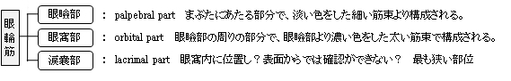
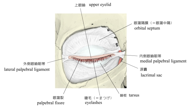
「 眼瞼部 ( Pars palpebralis ) は淡い色をした細い筋束からできており,眼瞼の実質のなかにあり,眼瞼縁の近くにまで及んでいる.これは外側では眼瞼の範囲を越えているが,上方と下方ではそれを越えていない.
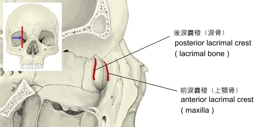
涙嚢部 ( pars sacci lacrimalis ) の線維は後涙嚢稜および涙嚢から起って,眼瞼に達する(図519,第II巻,眼の項参照).
眼窩部 ( Pars orbitalis ) はもはや眼瞼には属していない部分である.これは眼瞼部よりも厚くて,いっそう濃い色をしており,さらに太い筋束をもっている.上眼瞼の周りにある部分は扇形に広がり,しかもその程度も強いので,内側の線維はほとんど垂直に上方へすすみ眉の内側端部に達している.これが 眉頭下制筋 ( M. depressorcapitis supercilii ) であるが,一方ほかの線維は外側にあるものほどいっそう垂直からはなれた方向をとっている. 」
 |
|
|
|
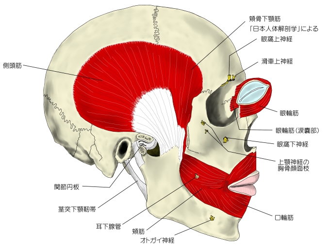
![]() 引 用 :「 日本人体解剖学 」
引 用 :「 日本人体解剖学 」
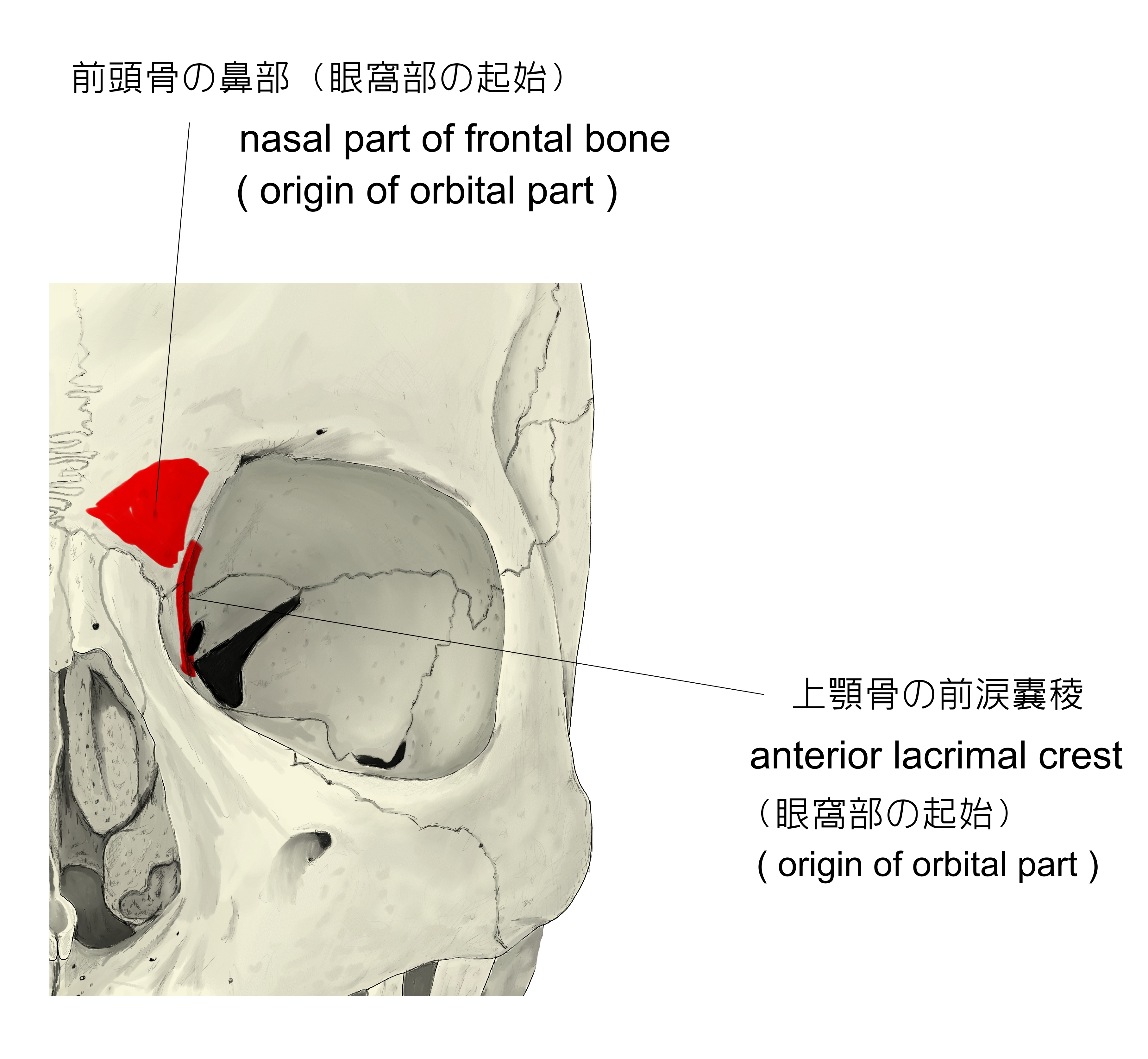
【 起 始 】: 眼瞼部 : 内側眼瞼靭帯
眼窩部 : 前頭骨 の 鼻部、上顎骨 の 前涙嚢稜、内側眼瞼靭帯
【 停 止 】: 眼瞼部 : 外側眼瞼靭帯
眼窩部 : 眼窩縁 を輪状に走り、一部は 外側眼瞼靭帯 あるいは眉の皮膚へ
涙嚢部 : 内眼角 で 眼瞼部 に移行する。
 |
 |
|
「 眼瞼を閉じ、( 眼瞼部、眼窩部 )、涙嚢を開ける。( 涙嚢部 )」 ( 日本人体解剖学 )
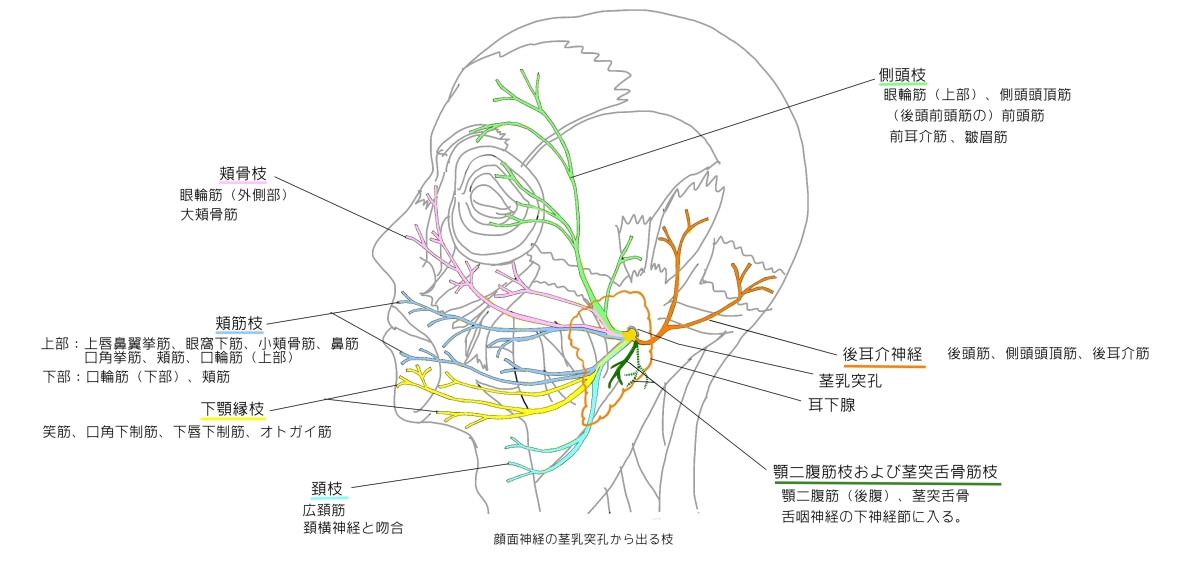
・ 神 経 : 顔面神経 の 側頭枝 および 頬骨枝 ( 日本人体解剖学 )
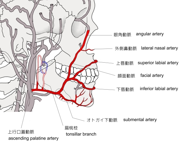
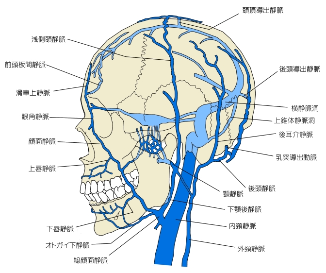
・ 脈 管 : 顔面動脈・静脈 の枝の 眼角動脈・静脈 またはその他あたりだと思われるが、詳しく言及している資料は見当たらない。
 |
 |
 |
The orbicularis oculi is a muscle in the face that closes the eyelids. It arises from the nasal part of the frontal bone, from the frontal process of the maxilla in front of the lacrimal groove, and from the anterior surface and borders of a short fibrous band, the medial palpebral ligament.
From this origin, the fibers are directed laterally, forming a broad and thin layer, which occupies the eyelids or palpebræ, surrounds the circumference of the orbit, and spreads over the temple, and downward on the cheek.
【 語 句 】
・ eyelid:まぶた ・nasal part of the frontal bone :前頭骨の ・frontal process of the maxilla :上顎骨の前頭突起 ・ lacrimal groove:涙嚢溝 ・ medial palpebral ligament:内側眼瞼靭帯 ・palpebræ :眼瞼 ・ circumference:周囲 ・ orbit:眼窩 ・ temple:こめかみ
There are at least 3 clearly defined sections of the orbicularis muscle. However, it is not clear whether the lacrimal section is a separate section, or whether it is just an extension of the preseptal and pretarsal sections.[1][2]
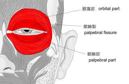
■ Orbital orbicularis 眼窩部 ■
The orbital portion is thicker and of a reddish color ; its fibers form a complete ellipse without interruption at the lateral palpebral commissure ; the upper fibers of this portion blend with the frontalis and corrugator.
■ Palpebral orbicularis 眼瞼部 ■
The palpebral portion of the muscle is thin and pale ; it arises from the bifurcation of the medial palpebral ligament, forms a series of concentric curves, and is inserted into the lateral palpebral raphe at the outer canthus (corner) of the eye.[3] The palpebral portion contains the preseptal and pretarsal muscles. The pretarsal orbicularis is thought to be responsible for the spontaneous blink
■ Lacrimal orbicularis 涙嚢部 ■
The lacrimal part is a small, thin muscle, about 6 mm in breadth and 12 mm in length, situated behind the medial palpebral ligament and lacrimal sac. It arises from the posterior crest and adjacent part of the orbital surface of the lacrimal bone, and passing behind the lacrimal sac, divides into two slips, upper and lower, which are inserted into the superior and inferior tarsi medial to the puncta lacrimalia ; occasionally it is very indistinct. The lacrimal orbicularis facilitates the tear pump into the lacrimal sac.[4]
【 語 句 】
・ pretarsal : 眼瞼前部の ・ reddish : 赤みがかった ・ ellipse : 楕円 ・ commissure : 交わり ・ frontalis : 前頭筋 ・ corrugator : 皺筋 ( 皺眉筋 (シュウビキン) のこと? ) ・ bifurcation : 分岐点 ・ concentric : 同心円をなして ・ lateral palpebral raphe : 外側眼瞼縫線 ・ outer canthus (corner) of the eye : 外眼角 ・ spontaneous : 自然に起こる ・ blink : まばたき ・ lacrimal sac : 涙嚢 ・ lacrimal bone : 涙骨 ・ puncta lacrimalia : 涙点? ・ indistinct : 不明瞭な ・ facilitate : 促進する、楽にする
The muscle acts to close the eye, and is the only muscle capable of doing so. Loss of function for any reason results in an inability to close the eye, necessitating eye drops at the minimum to surgical closure of the eye in extreme cases.
The palpebral portion acts involuntarily, closing the lids gently, as in sleep or in blinking ; the orbital portion is subject to conscious control. When the entire muscle is brought into action, the skin of the forehead, temple, and cheek is drawn toward the medial angle of the orbit, and the eyelids are firmly closed, as in photophobia. The skin thus drawn upon is thrown into folds, especially radiating from the lateral angle of the eyelids; these folds become permanent in senescence, and form the so-called "crow's feet". The Levator palpebræ superioris is the direct antagonist of this muscle; it raises the upper eyelid and exposes the front of the bulb of the eye. In addition, the orbital and palpebral portions can work independent of each other, as in the furrowing of the brows by contraction of the orbital to reduce glare while keeping the eyes open by virtue of the relaxation of the palpebral.[3]
【 語 句 】
・ necessitate : 必要とする ・ at the minimum : 最小限度 ・ surgical closure : 外科的縫合 ・ involuntarily : 意識しないで ・ photophobia: ・ photophobia : 羞明 (シュウメイ) ・radiate : 放出する、四方に広まる ・ senescence : 老齢 ・ Levator palpebrae superioris : 上眼瞼挙筋 ・ antagonist : 拮抗筋 ・ expose : 露出させる ・ bulb of the eye : 眼球 ・ furrow : しわを寄せる ・ glare : にらむ ・ by virtue of ~ : ~ のせいで
Each time the eyelids are closed through the action of the orbicularis, the medial palpebral ligament is tightened, the wall of the lacrimal sac is thus drawn lateralward and forward, so that a vacuum is made in it and the tears are sucked along the lacrimal canals into it. The lacrimal part of the orbicularis oculi draws the eyelids and the ends of the lacrimal canals medialward and compresses them against the surface of the globe of the eye, thus placing them in the most favorable situation for receiving the tears; it also compresses the lacrimal sac. This part comprises two pieces : Horner's muscle and the muscle of Riolan, the latter helps hold the eyelids together to keep the lacrimal passage waterproof.[3]
Associated pathology, such as a lesion of the facial nerve seen in Bell's palsy results in the inability to blink or close the ipsilateral eyelid. Subsequent lack of irrigation increases the risk of corneal inflammation and ulcers.[citation needed]
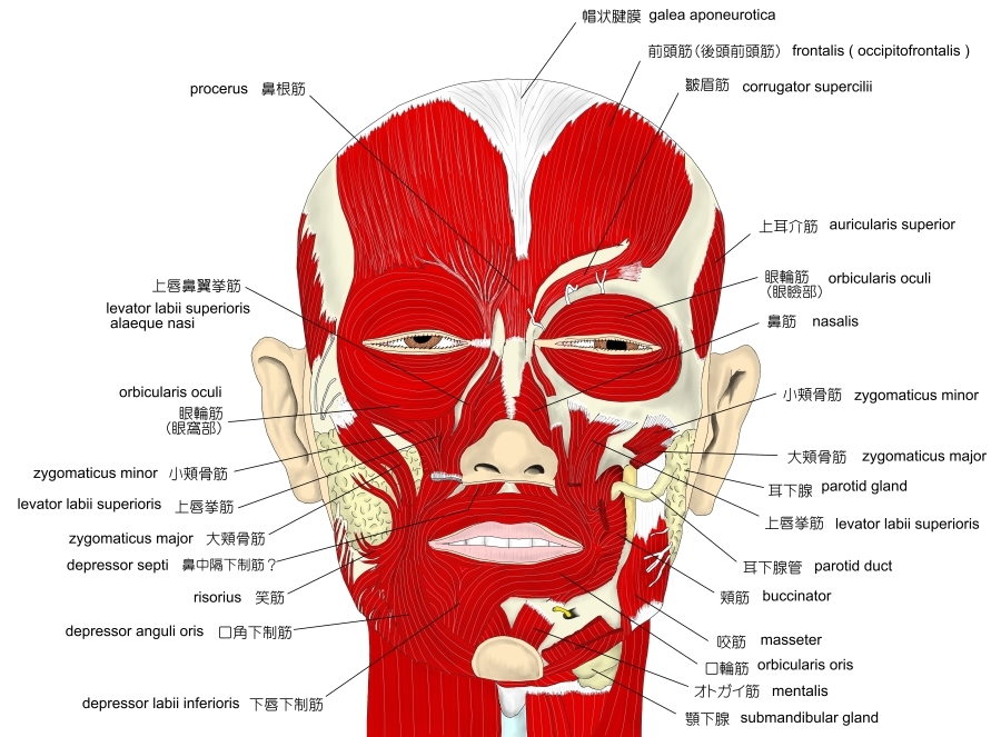
A number of auxiliary muscles assist in cooperating with the eyelid muscles. For example, the corrugator supercilii pulls the eyebrows to the bridge of the nose, making a roof over the middle of the forehead and forehead wrinkles, used mainly to protect the eyes from excess sunlight. The procerus ( pyramidalis ) muscles, in the bridge of the nose, arise from the lower nasal bone to the lower forehead, on each side of the midline. The procerus muscles pull the skin into horizontal wrinkles. The frontalis muscle, which runs from the upper forehead, halfway between the coronal suture ( which traverses the top of the skull ) and the top edge of the orbit, attaches to the eyebrow skin. Since it pulls the eyebrows upward, it is the antagonist of the orbicularis oculi. It is used in looking up, and increasing vision if there is insufficient light or when objects are far away.[3]
【 語 句 】
・ lacrimal canals : 涙管 ・ comprise : 構成する ・ lacrimal passage : 涙道 ・ pathology : 病理学 ・ lesion : 傷害 ・ Bell's palsy : ベル麻痺? ・ipsilateral : 同側性の ・subsequent : 続いて起こる ・ irrigation : 潅水 ・ corneal : 角膜の ・ inflammation : 炎症 ・ ulcer : 潰瘍 ・ auxiliary : 予備の ・ corrugator supercilii : 皺眉筋 ・ bridge of the nose : 鼻梁 ・ wrinkles : 小じわ ・ excess : 過度な ・ procerus : 鼻根筋 ・ coronal suture : 冠状縫合