

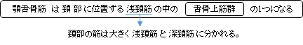
顎舌骨筋( がくぜっこつきん、英:mylohyoid muscle )
・ 概 要 |
・ 作 用 |
・ イラスト掲載サイ |
|
・ イラスト |
・ 神経 / 脈管 |
||
・ 起始 / 停止 |
・ Wikipedia |
「 日本人体解剖学 」には以下のような解説文が見られる。
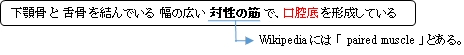
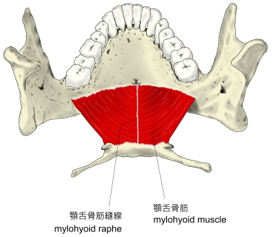
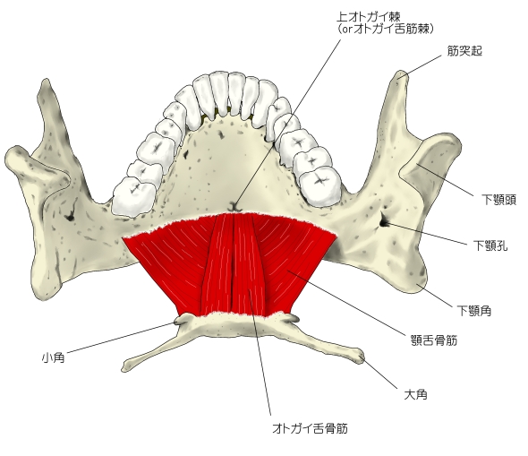
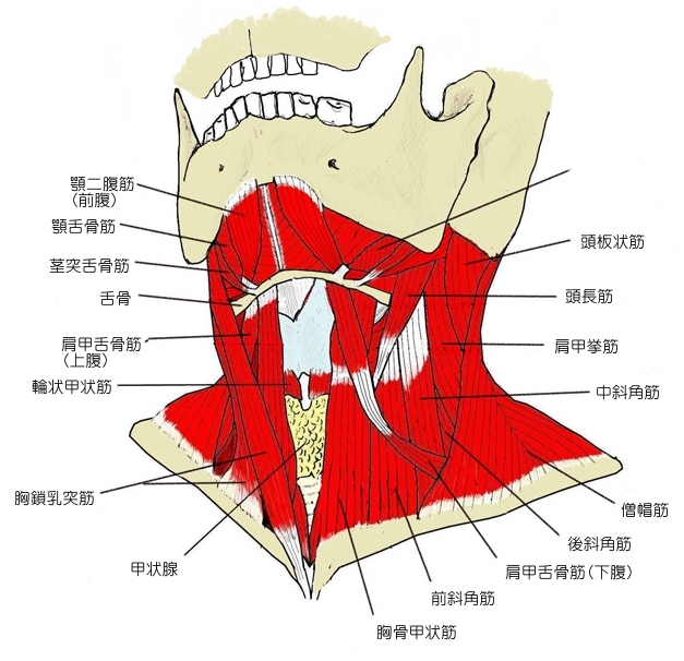
「 左右の両筋は、全体として平たい三角形となり、口腔底を作り舌を乗せているので 口隔膜 とも呼ばれる。」
 |
 |
 |
 |
 |
 |
 |
起始部より内側下方に走る。
 |

|
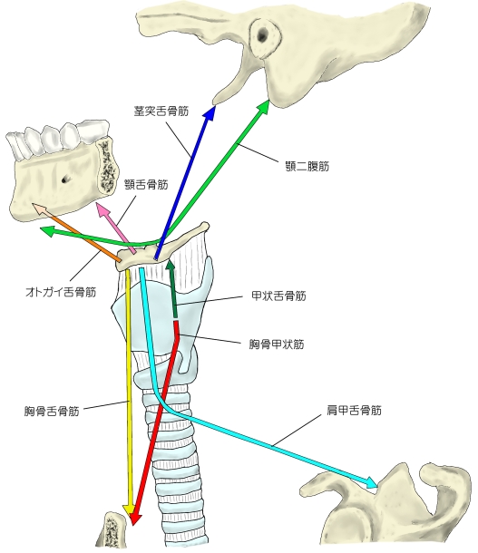
「 下顎骨を固定すると舌骨を挙上し、舌骨を固定すると下顎骨を引き下げる。」 ( 日本人体解剖学 )
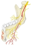
・ 神 経 : 顎舌骨筋神経 ( 下顎神経( 三叉神経の第3枝 )の枝の 下歯槽神経 の枝 )
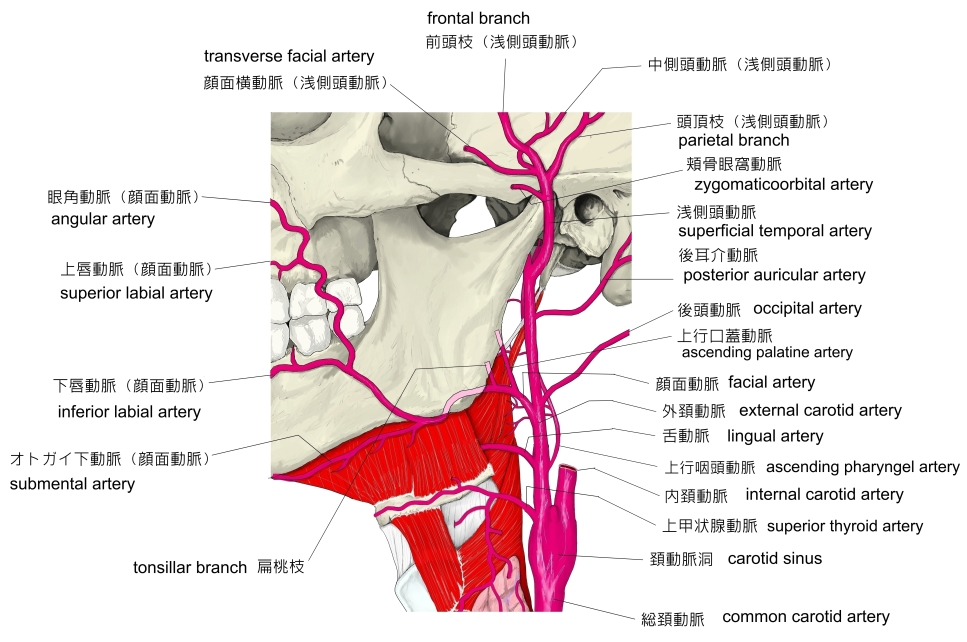
・ 動 脈 : オトガイ下動脈 ( 顔面動脈 の枝 )あたりだと思われるが、詳細は不明。
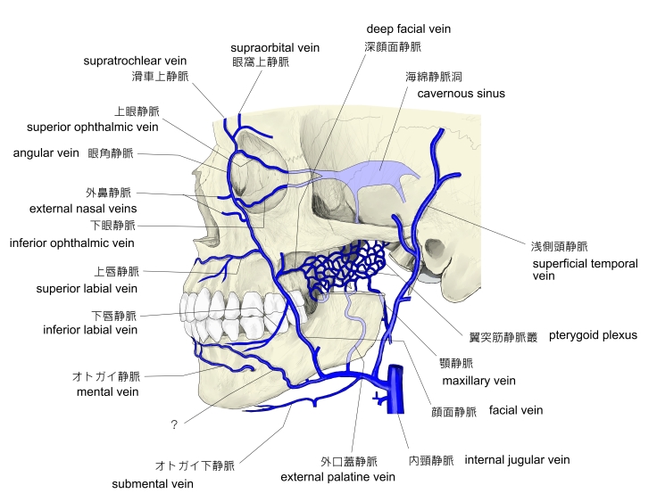
・ 静 脈 : もし血液を供給する動脈が オトガイ下動脈 であれば、静脈は オトガイ下静脈 になると思われる
が、詳細は不明。
|
|
|
The mylohyoid muscle is a paired muscle running from the mandible to the hyoid bone, forming the floor of the oral cavity ( the mouth ).[1] :987 It is named after its two attachments near the molar teeth ( "mylo" comes from the Greek word for "molar" ).[2] These muscles are mesodermal in embryologic origin. The mylohyoid muscle is derived from the first pharyngeal arch.
【 Structure 】
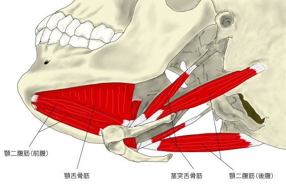
The mylohyoid muscle is flat and triangular, and is situated immediately superior to the anterior belly of the digastric muscle. It is classified as an anterior suprahyoid muscle. [ clarification needed ] Together, the paired mylohyoid muscles form a muscular floor ( diaphragm ) for the oral cavity (the mouth).[3] :212
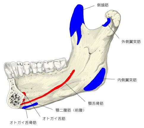
The two mylohyoid muscles arise from the mandible at the mylohyoid line, which extends from the mandibular symphysis in front to the last molar tooth behind. The posterior fibers pass inferomedially and insert at anterior surface of the hyoid bone. The medial fibres of the two mylohyoid muscles unite in a midline raphe (where two muscles intermesh).[1] :987-8
The mylohyoid muscle separates the sublingual space from the submandibular space, which communicate in a lateral gap between the mylohyoid and geniohyoid muscles.[4] The submandibular gland wraps around the edges of the mylohyoid, and is divided into superficial and deep lobes above and below the muscle.[1] :997
【 語 句 】
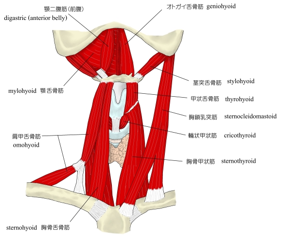
・ mandible : 下顎骨 ・ hyoid bone : 舌骨 ・ oral cavity : 口腔 ・ molar teeth : 大臼歯 ・ mesodermal : 中胚葉性の ・ embryologic : 発生学的な ・ be derived from ~ : ~ に由来する? ・ pharyngeal arch : 咽頭弓 ・ anterior belly of the digastric muscle : 顎二腹筋の前腹 ・ suprahyoid muscle : 舌骨上筋 ・ diaphragm : 隔膜 ・ sublingual space : 舌下隙 ・ submandibular space : 顎下隙 ・ geniohyoid muscle : オトガイ舌骨筋 ・ submandibular gland : 顎下腺
【 Innervation 】
The mylohyoid muscle is innervated by a branch of the mandibular nerve, the inferior alveolar nerve. A specific branch of this nerve, the mylohyoid nerve, emerges to give motor supply to the muscle.[1] :987
【 Variations 】
The mylohyoid may be united to or replaced by the anterior belly of the digastric muscle ; accessory slips to other hyoid muscles are frequent. This median raphé is sometimes absent ; the fibers of the two muscles are then continuous. [citation needed]
An area of herniation of the sublingual gland, blood vessels, or fat, may be present, with studies reporting this in 10-50% of people.[4]
【 Function 】
The mylohyoid elevates the hyoid and the tongue. This is particularly important during swallowing and speaking. Alternatively, if other muscles are used to keep the position of the hyoid fixed, then the mylohoid depresses the mandible.[1] :987 It also functions as reinforcing the floor of mouth.[1] :987
【 語 句 】
・ mandibular nerve : 下顎神経 ・ inferior alveolar nerve : 下歯槽神経 ・ mylohyoid nerve : 顎舌骨筋神経 ・ herniation : ヘルニア形成 ・ sublingual gland : 舌下腺 ・ reinforce : 補強する
![]()