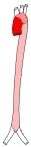
大動脈弓とは
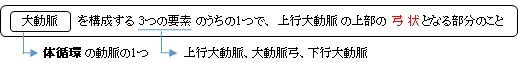
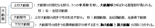
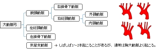
以下は大動脈の構成を簡単に表したものとなる。
以下は「 日本人体解剖学 」の解説をまとめたものとなる。
・ 頂点の高さは第一肋骨の胸骨付着部の上縁にあたる。
・ 大動脈弓は縦隔胸膜と肺によって覆われている。
・ 気管支分岐部のところを後方へ超えていく。
・ 後方では右側に食道が位置する。
・ 上縁には左腕頭静脈が隣接し、その下部には右肺動脈が左から右に走り、左反回神経 が前から後ろに走る。
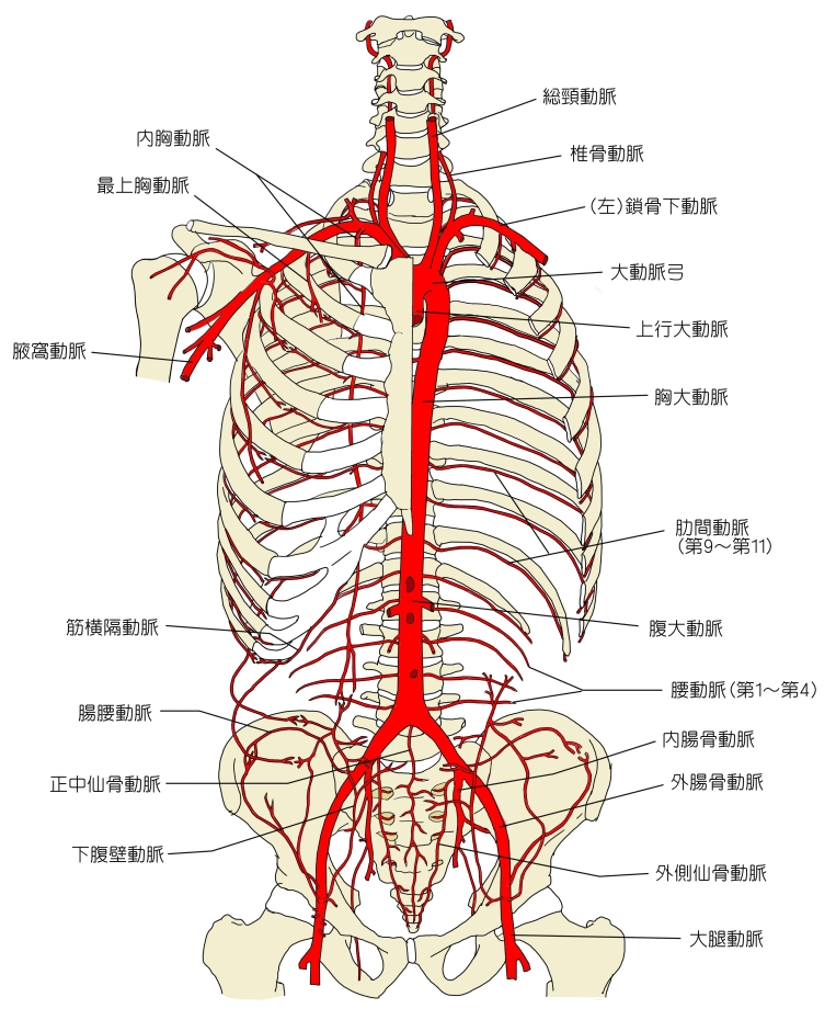
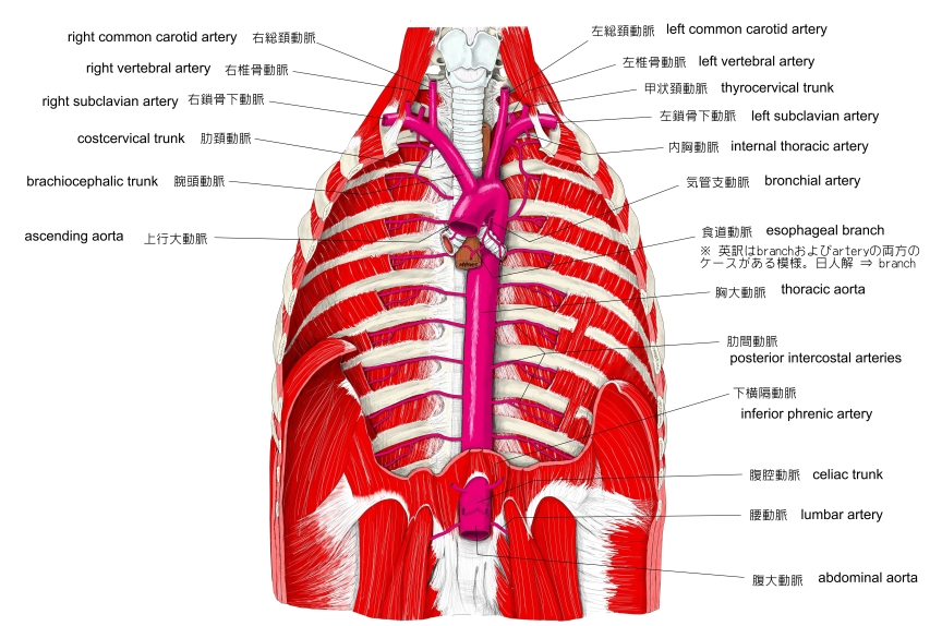
上半身の主な動脈
|
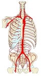
胸部腹部の
|
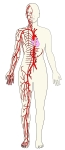
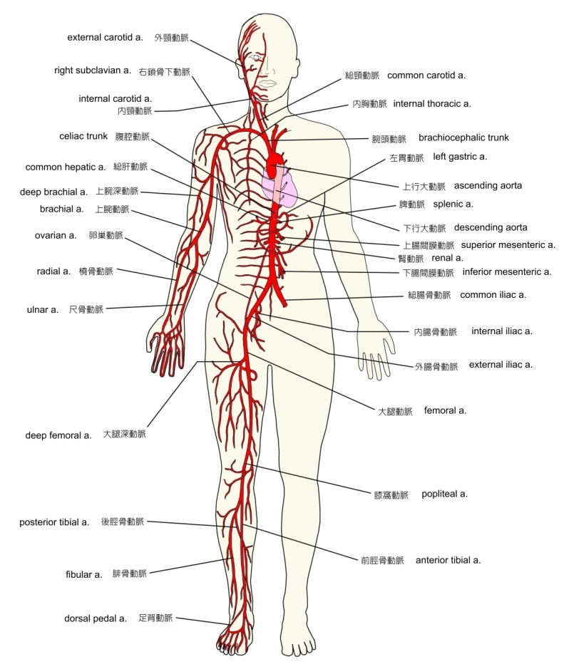
体循環の主な動脈
|
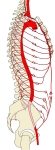
大動脈
|
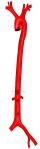
大動脈 |
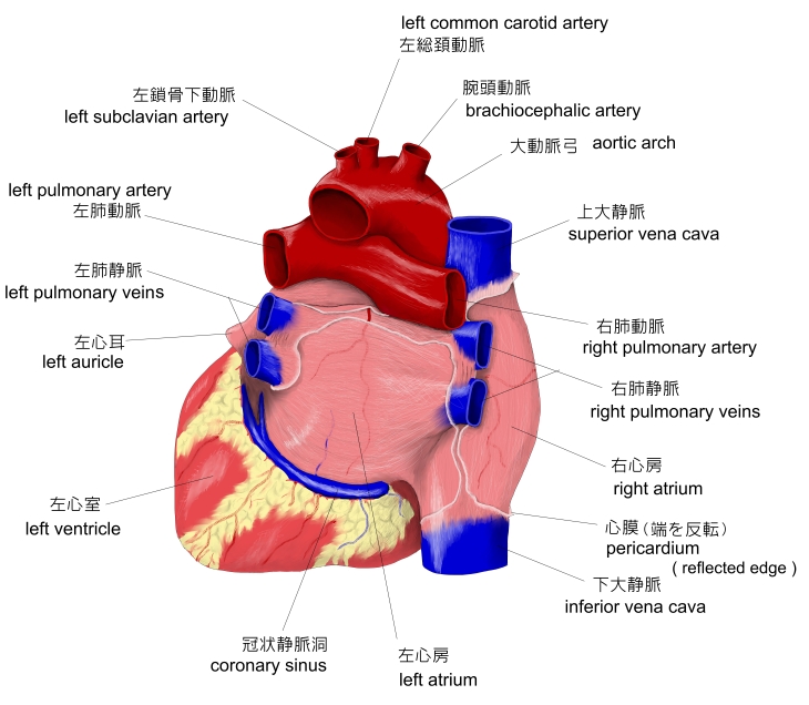
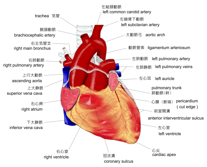
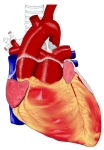
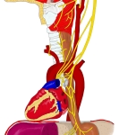
心臓(前面)
|
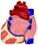
心臓(後面)
|
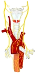
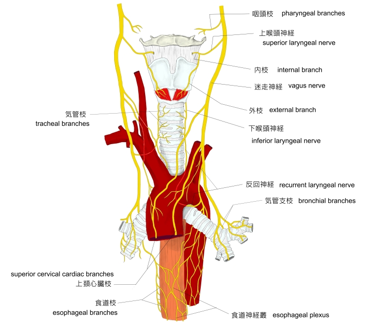
迷走神経
|
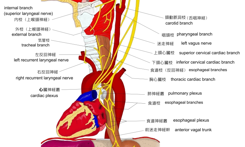
迷走神経一部
模型図
|
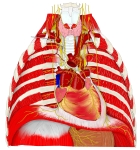
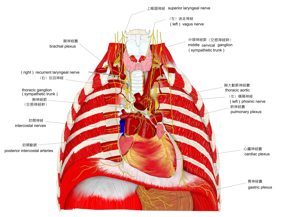
胸腔の神経
、筋など
|
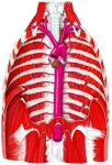
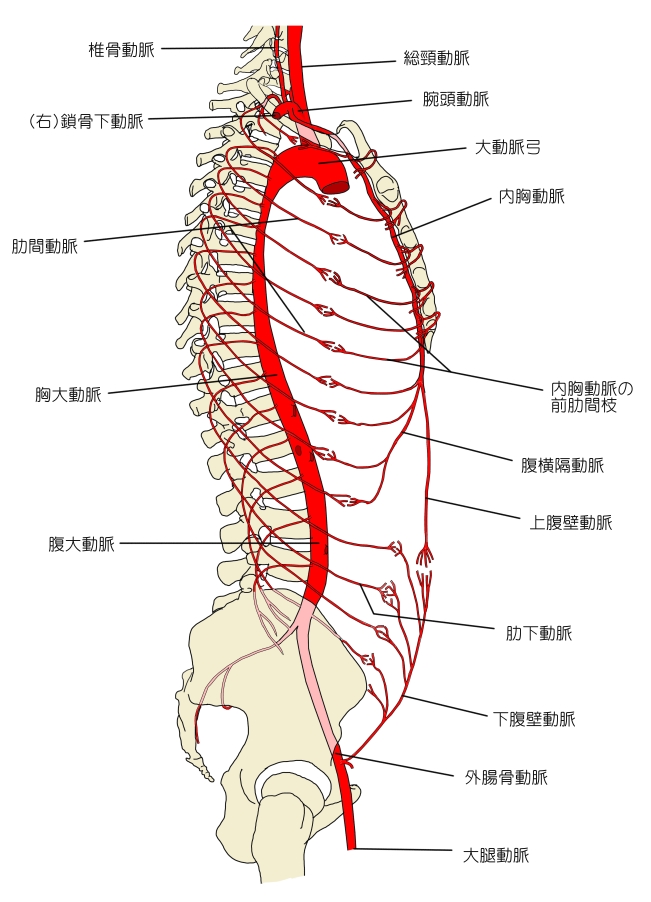
胸大動脈とその枝
|
|

・ しばしば1~2本の気管支動脈が起こることがある。
・「 稀には腕頭動脈と左総頚動脈が共通幹をもって起こることがあり、また左椎骨動脈が直接大動脈弓から出ることなどがある。」( 日本人体解剖学 )


■ 異 常 ■
「 日本人体解剖学 」には以下のような解説文が見られる。
「 大動脈弓の高さの異常 : 大動脈弓の高さが正常よりも高く、まれに胸骨柄の上縁に達することがある。また、正常よりも低く、はなはだしい時は胸骨柄の上縁から6~7㎝も下方にあることもある。
大動脈弓の左曲がり : 心臓その他の内臓の位置が反対(内臓逆位)になっている結果に生じ、また大動脈弓の位置には変化がなく左腕頭動脈・右総頚動脈・右鎖骨下動脈が出ている場合がある。 」

以下は「 Wikipedia 」の解説文となる。
「 The aortic arch, arch of the aorta, or transverse aortic arch (English: /e????rt?k/[1][2]) is the part of the aorta between the ascendingand descending aorta. The arch travels backward, so that it ultimately runs to the left of the trachea.
【 語 句 】
・ ultimately : 最後に ・ trachea : 気管
【 structure 】
At the cellular level, the aorta and the aortic arch are composed of three layers: The tunica intima, which surrounds the lumen and is composed of simple squamal epithelial cells; the tunica media, composed of smooth cell muscles and elastic fibers; and, the tunica adventitia, composed of loose collagen fibers.[3] Innervated by barometric nerve terminals, the aortic arch is responsible for sensing changes in the dilation of the vascular walls, inducing changes in heart rate to compensate for changes in blood pressure.[4]
【 語 句 】
・ tunica intima : 内膜 ・ epithelial cell : 上皮細胞 ・ tunica adventitia : 外膜 ・ tunica media : 中膜
・ nerve terminal : 神経終末 ・ dilation : 拡張 ・ compensate : 償う
The aorta begins at the level of the upper border of the second sternocostal articulation of the right side, and runs at first upward, backward, and to the left in front of the trachea; then travels backward on the left side of the trachea and finally passes downward on the left side of the body of the fourth thoracic vertebra.[5] At this point the aortic arch continues as the descending aorta.[5]:214[6]
The aortic arch has three branches. The first, and largest, branch of the arch of the aorta is the brachiocephalic trunk, which is to the right and slightly anterior to the other two branches and originates behind the manubrium of the sternum. Next, the left common carotid artery originates from the aortic arch to the left of the brachiocephalic trunk, then ascends along the left side of the trachea and through the superior mediastinum. Finally, the left subclavian artery comes off of the aortic arch to the left of the left common carotid artery and ascends, with the left common carotid, through the superior mediastinum and along the left side of the trachea.[7]:216 An anatomical variation is that the left vertebral artery can arise from the aortic arch instead of the left subclavian artery.
【 語 句 】
・ manubrium : 胸骨柄 ・ mediastinum : 縦隔 ・ vertebral artery : 積骨動脈
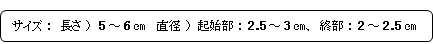
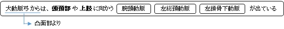
The arch of the aorta forms two curvatures: one with its convexity upward, the other with its convexity forward and to the left. Its upper border is usually about 2.5 cm. below the superior border to the manubrium sterni.[5] Blood flows from the upper curvature to the upper regions of the body, located above the heart - namely the arms, neck, and head.
Coming out of the heart, the thoracic aorta has a maximum diameter of 40 mm at the root. By the time it becomes the ascending aorta, the diameter should be < 35–38 mm, and 30 mm at the arch. The diameter of the descending aorta should not exceed 25 mm.[8][9] The arch of the Aorta lies within the mediastinum.
【 語 句 】
・ convexity : 凸面
【 development 】
The aortic arch is the connection between the ascending and descending aorta, and its central part is formed by the left 4th aortic arch during early development.[10]
The ductus arteriosus connects to the lower part of the arch in foetal life. This allows blood from the right ventricle to mostly bypass the pulmonary vessels as they develop.
The final section of the aortic arch is known as the isthmus of aorta. This is so called because it is a narrowing (isthmus) of the aorta as a result of decreased blood flow when in foetal life. As the left ventricle of the heart increases in size throughout life, the narrowing eventually dilates to become a normal size. If this does not occur, this can result in coarctation of the aorta.[11][12] The ductus arteriosus connects to the final section of the arch in foetal life and the ligamentum arteriosum when the ductus arteriosus regresses.[11]
【 語 句 】
・ ductus arteriosus : 動脈管 ・ foetal : 胎児の ・ right ventricle : 右心室 ・ isthmus of aorta : 大動脈峡部
・ coarctation : 狭窄 ・ regress : 退化する
【 variation 】
There are three common variations in how arteries branch from the aortic arch. In about 75% of individuals, the branching is "normal", as described above. In some individuals the left common carotid artery originates from the brachiocephalic artery rather than the aortic arch. In others, the brachiocephalic artery and left common carotid artery share an origin.[13] This variant is found in approximately a 20% of the population. In a third variant, the brachiocephalic artery splits into three arteries: the left common carotid artery, the right common carotid artery and the right subclavian artery; this variant is found in an estimated 7% of individuals. 」
【 イラスト掲載サイト 】
・ イラストや写真を掲載しているサイト-Ⅰ
・ イラストや写真を掲載しているサイト-Ⅱ
・ イラストや写真を掲載しているサイト-Ⅲ
・ イラストや写真を掲載しているサイト-Ⅳ
・ イラストや写真を掲載しているサイト-Ⅴ

|