【概 要】
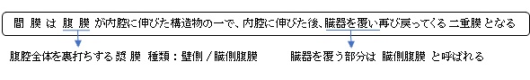
・間膜は発生学的に腹側間膜と背側間膜に分けられる。
・部位ごとに個別の名称がつけられている。 例)腸間膜、横行結腸膜など
以下は腹膜が内腔に伸びた構造物の簡単な表となる。


まとめ
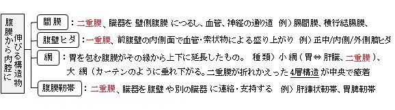
【臓器との関係性】
間膜と臓器との関係性は以下の2種類に分けることができる。
■ 臓器を包み込むタイプ ■
|
臓 器 |
間膜名 |
可動性 |
備 考 |
1 |
|
|
◎ 非常に高い |
最も典型的な「間膜臓器」 |
2 |
|
|
◎ 高い |
末端部はやや制限される |
3 |
|
|
○ 中等度 |
胃()と強く関係する |
4 |
|
|
◎ 高い |
捻転しやすい(臨床的に重要) |
5 |
|
|
△ 低〜中 |
位置の個体差が極端 |
■ 臓器の一部だけを覆うタイプ ■
このタイプは次の臓器が挙げられ、以下の特徴を有する。
| |
臓 器 |
間膜名 |
可動性 |
備 考 |
1 |
|
|
△ 低い |
|
2 |
|
|
◎ 高い |
本来「包み込むタイプ」だが、 小網・ 小網で固定される |
3 |
|
特になし |
×殆どない |
|
・他の臓器と連結する。
・後腹壁に直接つながらない。(消えたのではなく、再配置・融合して見えなくなった。)
・発生学的に「胃の間膜由来」
間膜が臓器同士をつなぐ理由
→ もともと壁側腹膜由来の間膜が発生中に移動・癒着した結果で、妊娠4か月(16週)頃にはほぼ現在の配置に近い状態になる。
👉 「間膜+腹膜ヒダの中間的存在」
🟢 このタイプに属する臓器(すべて)
① 胃まわり
② 脾臓
③ 結腸との連結
👉 まとめると、このタイプの中心は:
✅ 胃
✅ 脾臓
(+横行結腸との連結部)
■ 種 類 ■
以下が主な間膜になる。
消化管 |
1 |
|
|
2 |
|
|
3 |
|
|
4 |
|
|
胃 |
1 |
|
|
2 |
|
|
3 |
|
|
4 |
|
|
5 |
|
|
肝臓 |
1 |
|
|
2 |
|
|
3 |
|
|
生殖器 |
1 |
子宮広間膜 |
子宮全体を骨盤壁につなぐ。 |
2 |
卵巣間膜 |
卵巣を広間膜につなぐ。 |
3 |
卵管間膜 |
卵管を広間膜につなぐ。 |
他 |
1 |
|
|
・胎生期には上行結腸間膜、下行結腸間膜というものも見られる。
【参考となるサイト】
以下は「日本人体解剖学 (下巻) 」の「腹膜」の項の「間膜」の解説文となる。
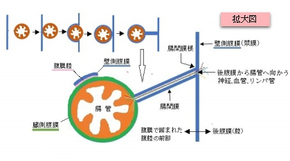
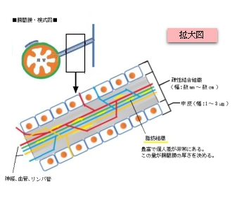
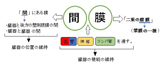
「腹膜内臓器である空腸や回腸のように腹壁から離れて存在する臓器の場合には、壁側腹膜は腹壁から臓器の表面に達し、臓器の表面をおおったのち再び腹壁に返るため、往復2枚の腹膜は合して間膜と呼ばれる二重の膜をつくる。この中を血管、リンパ管、神経が通る。間膜はそれぞれの臓器に従い名称がつけられ、たとえば空腸と回腸では腸間膜、S状結腸ではS状結腸間膜という。 」
「Wikipedia」では以下のように解説している?
In human anatomy, the mesentery is an organ that attaches the intestines to the posterior abdominal wall, consisting of a double fold of the peritoneum. It helps (among other functions) in storing fat and allowing blood vessels, lymphatics, and nerves to supply the intestines.
The mesocolon (the part of the mesentery that attaches the colon to the abdominal wall) was formerly thought to be a fragmented structure, with all named parts—the ascending, transverse, descending, and sigmoid mesocolons, the mesoappendix, and the mesorectum—separately terminating their insertion into the posterior abdominal wall.[2] However, in 2012, new microscopic and electron microscopic examinations showed the mesocolon to be a single structure derived from the duodenojejunal flexure and extending to the distal mesorectal layer.[2][3] Thus the mesentery is an internal organ.
【Structure 】
The mesentery of the small intestine arises from the root of the mesentery (or mesenteric root) and is the part connected with the structures in front of the vertebral column. The root is narrow, about 15 cm long, 20 cm in width, and is directed obliquely from the duodenojejunal flexure at the left side of the second lumbar vertebra to the right sacroiliac joint. The root of the mesentery extends from the duodenojejunal flexure to the ileocaecal junction. This section of the small intestine is located centrally in the abdominal cavity and lies behind the transverse colon and the greater omentum.
The mesentery becomes attached to the colon at the gastrointestinal margin and continues as the several regions of the mesocolon. The parts of the mesocolon take their names from the part of the colon to which they attach. These are the transverse mesocolon attaching to the transverse colon, the sigmoid mesocolon attaching to the sigmoid colon, the mesoappendix attaching to the appendix, and the mesorectum attaching to the upper third of the rectum.
The mesocolon regions were traditionally taught to be separate sections with separate insertions into the posterior abdominal wall. In 2012, the first detailed observational and histological studies of the mesocolon were undertaken and this revealed several new findings.[6] The study included 109 patients undergoing open, elective, total abdominal colectomy. Anatomical observations were recorded during the surgery and on the post-operative specimens.
These studies showed that the mesocolon is continuous from the ileocaecal to the rectosigmoid level. It was also shown that a mesenteric confluence occurs at the ileocaecal and rectosigmoid junctions, as well as at the hepatic and splenic flexures and that each confluence involves peritoneal and omental attachments. The proximal rectum was shown to originate at the confluence of the mesorectum and mesosigmoid. A plane occupied by perinephric fascia was shown to separate the entire apposed small intestinal mesentery and the mesocolon from the retroperitoneum. Deep in the pelvis, this fascia coalesces to give rise to presacral fascia.