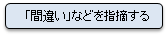
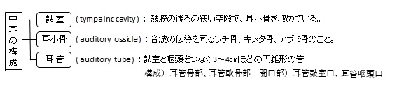
以下は中耳の構成要素を簡単に表した図となる。
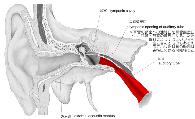
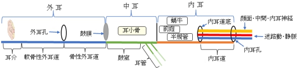
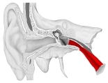
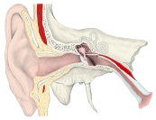
以下は外耳から内耳にかけての簡単な図となる。

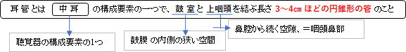
以下、耳管の特徴を簡単に記す。
・粘膜は咽頭鼻部を覆う粘膜の続きとなる。
・多列線毛柱上皮からでき、線毛運動は咽頭方に向かっている。
・粘膜固有層は結合組織からでき、上皮と骨膜あるいは軟骨膜とを結合している。
・耳管腺は粘液腺で、特に軟骨部に多量に存在している。
・耳管扁桃:リンパ小節(耳管リンパ小節)は咽頭への開口部である耳管咽頭口に多く、 耳管扁桃をつくる。
「日本人体解剖学 (下巻) 」には以下のような解説が見られる。
「耳管は、通常、特に耳管峡においてほとんど閉ざされたようになっているが(安全管を除き)、嚥下運動をすると口蓋帆挙筋の収縮によって開き空気が鼓室に出入する。これによって鼓膜の内外、すなわち鼓室と外耳道との気圧の均衡を保ち、音響の内耳に伝わる作用を助ける。小児、ことに新生児では耳管峡は見形成で、また耳管が成人よりもいっそう水平位に近いため、鼻腔または咽頭の印象が鼓膜に波及しやすい。」
 |
 |
 |
 |
右耳・冠状断面・前面 |
右耳・冠状断面・前面 |
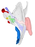
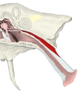
右側頭骨・上面 |
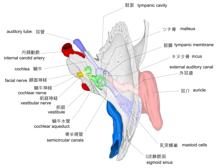
右耳管・前面 |
 |
|
|
|
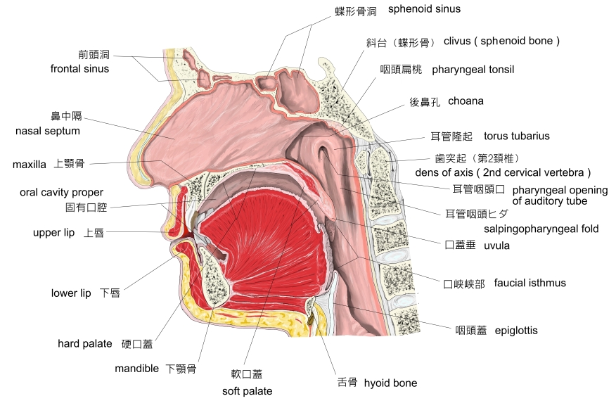
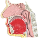
正中断面・左より見て
|
|
|
|

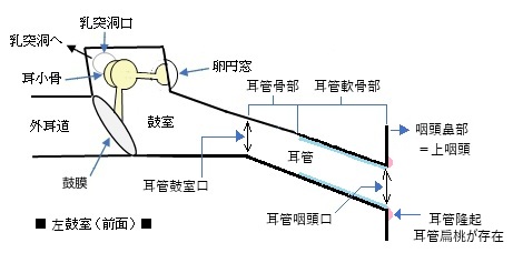
起 始 : 耳管鼓室口 (tympanic opening of auditory tube) : 鼓室の頚動脈壁(前壁)にある口
走 行 : 側頭骨の筋耳管菅の下部である耳管半管の中を走る。蝶錐体裂のあたりで軟骨部に移行する。
形 状 : 管径の割に比較的広い円みをおびた三角形の腔となる。
耳管蜂巣 (tubal air cells) : 骨部には腺がなく、耳管鼓室口の近くには数個の小さな粘膜の陥入があり、これを耳管蜂巣という。「日本人体解剖学 (下巻) 」
耳管峡 (isthmus of auditory tube) : 骨部と軟骨部の移行部のことで、耳管の中で最も狭い腔となっているところ。
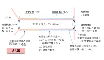

概要:耳管峡から耳管咽頭口に至るまでの間で、ほぼ全体として樋状(半管状)を呈する。
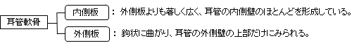
区別:耳管軟骨を内側板と外側板とに区別する。
|
|
|
右耳・冠状断面・前面
|
右側頭骨・上面 |
右耳管・前面
|

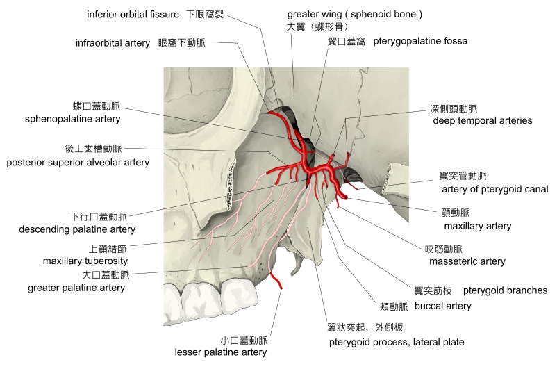
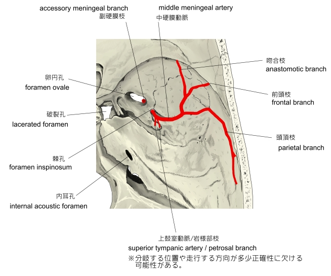
・血 管 : 翼突管動脈、上行咽頭動脈、中硬膜動脈の枝がくる。
・リンパ管 : 咽頭後リンパ節に入る。
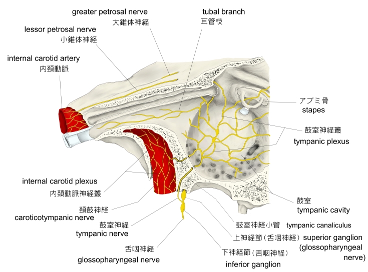
・神 経 : 鼓室神経叢の耳管枝および咽頭神経叢の枝。

In anatomy, the Eustachian tube, also known as the auditory tube or pharyngotympanic tube,[1] is a tube that links the nasopharynx to the middle ear, of which it is also a part. In adult humans, the Eustachian tube is approximately 35 mm (1.4 in) long and 3 mm (0.12 in) in diameter.[2] It is named after the sixteenth-century Italian anatomist Bartolomeo Eustachi.[3]
In humans and other land animals, both the middle ear and the ear canal are normally filled with air. Unlike the air of the ear canal, however, the air of the middle ear is not in direct contact with the atmosphere outside the body; thus, a pressure difference can develop between the atmospheric pressure of the ear canal and the middle ear. Normally, the Eustachian tube is collapsed, but it gapes open with swallowing and with positive pressure, allowing the middle ear's pressure to adjust to the atmospheric pressure. When taking off in an aircraft, the ambient air pressure goes from higher (on the ground) to lower (in the sky). The air in the middle ear expands as the plane gains altitude, and pushes its way into the back of the nose and mouth; on the way down, the volume of air in the middle ear shrinks, and a slight vacuum is produced. Active opening of the Eustachian tube (through actions like swallowing or the Valsalva maneuver) is required to equalize the pressure between the middle ear and the ambient atmosphere as the plane descends. A diver also experiences this change in pressure, but with greater rates of pressure change; active opening of the Eustachian tube is required more frequently as the diver goes deeper, into higher pressure.
【 語 句 】
・ : ・ : ・ : ・ : ・ : ・ : ・ : ・ : ・ : ・ : ・ : ・ : ・ : ・ : ・ : ・ :
The Eustachian tube extends from the anterior wall of the middle ear to the lateral wall of the nasopharynx, approximately at the level of the inferior nasal concha. It consists of a bony part and a cartilaginous part.
Bony part[edit]
The bony part (1⁄3) nearest to the middle ear is made of bone and is about 12 mm in length. It begins in the anterior wall of the tympanic cavity, below the septum canalis musculotubarius, and, gradually narrowing, ends at the angle of junction of the squamous and the petrous parts of the temporal bone, its extremity presenting a jagged margin which serves for the attachment of the cartilaginous part.[5] The vestibule of the Eustachian tube is known as the protympanum,[6] The protympanum is also known as the anterior part of the bony part of the tube.[7]
Cartilaginous part[edit]
The cartilaginous part of the Eustachian tube is about 24 mm in length and is formed of a triangular plate of elastic fibrocartilage, the apex of which is attached to the margin of the medial end of the bony part of the tube, while its base lies directly under the mucous membrane of the nasal part of the pharynx, where it forms an elevation, the torus tubarius or cushion, behind the pharyngeal opening of the auditory tube.
The upper edge of the cartilage is curled upon itself, being bent laterally so as to present on transverse section the appearance of a hook; a groove or furrow is thus produced, which is open below and laterally, and this part of the canal is completed by fibrous membrane. The cartilage lies in a groove between the petrous part of the temporal bone and the great wing of the sphenoid; this groove ends opposite the middle of the medial pterygoid plate. The cartilaginous and bony portions of the tube are not in the same plane, the former inclining downward a little more than the latter. The diameter of the tube is not uniform throughout, being greatest at the pharyngeal opening, least at the junction of the bony and cartilaginous portions, and again increased toward the tympanic cavity; the narrowest part of the tube is termed the isthmus.
The position and relations of the pharyngeal opening are described with the nasal part of the pharynx. The mucous membrane of the tube is continuous in front with that of the nasal part of the pharynx, and behind with that of the tympanic cavity; it is covered with ciliated pseudostratified columnar epithelia and is thin in the osseous portion, while in the cartilaginous portion it contains many mucous glands and near the pharyngeal orifice a considerable amount of adenoid tissue, which has been named by Gerlach the tube tonsil.
【 語 句 】
・ : ・ : ・ : ・ : ・ : ・ : ・ : ・ : ・ : ・ : ・ : ・ : ・ : ・ : ・ : ・ :
Muscles[edit]
There are four muscles associated with the function of the Eustachian tube:
The tube is opened during swallowing by contraction of the tensor veli palatini and levator veli palatini, muscles of the soft palate.[1]
New anatomical perspectives[edit]
More recently, two developments have enhanced our understanding of the anatomy of the eustachian tube: Valsalva computerized tomography and endoscopic ear surgery.[9]
- Given the greater access to the ear anatomy using endoscopic methods, it has been suggested that the bony part of the eustachian tube is really the anterior extension of the middle ear cavity, or the "Protympanum". The term "Eustachian Tube" should be limited to the fibrocartilaginous structure connecting the protympanum to the nasopharynx.[4]
- The Eustachian tube is a sac like irregular structure rather than a tubular structure.
- The ear side of the eustchian tube is by far the narrowest segment, called isthmus, and is probably the site of possible obstructive pathology causing chronic ear disease.[10]
The Eustachian tube is derived from the dorsal part of the first pharyngeal pouch and second endodermal pouch, which during embryogenesis forms the tubotympanic recess. The distal part of the tubotympanic sulcus gives rise to the tympanic cavity, while the proximal tubular structure becomes the Eustachian tube. It helps transformation of sound waves.
【 語 句 】
・ : ・ : ・ : ・ : ・ : ・ : ・ : ・ : ・ : ・ : ・ : ・ : ・ : ・ : ・ : ・ :
Pressure equalization[edit]
Under normal circumstances, the human Eustachian tube is closed, but it can open to let a small amount of air through to prevent damage by equalizing pressure between the middle ear and the atmosphere. Pressure differences cause temporary conductive hearing loss by decreased motion of the tympanic membrane and ossicles of the ear.[11] Various methods of ear clearing such as yawning, swallowing, or chewing gum may be used to intentionally open the tube and equalize pressures. When this happens, humans hear a small popping sound, an event familiar to aircraft passengers, scuba divers, or drivers in mountainous regions. Devices assisting in pressure equalization include an ad hoc balloon applied to the nose, creating inflation by positive air pressure.[12] Some people learn to voluntarily 'click' their ears, together or separately, performing a pressure equalizing routine by opening their Eustachian tubes when pressure changes are experienced, as in ascending/descending in aircraft, mountain driving, elevator lift/drops, etc. Some are even able to deliberately keep their Eustachian tubes open for a brief period, and even increase or decrease air pressure in the middle ear. The 'clicking' can actually be heard by putting one's ear to another's while performing the clicking sound. This voluntary control may be first discovered when yawning or swallowing, or by other means (above). Those who develop this ability may discover that it can be done deliberately without force even when there are no pressure issues involved.
Mucus drainage[edit]
The Eustachian tube also drains mucus from the middle ear. Upper respiratory tract infections or allergies can cause the Eustachian tube, or the membranes surrounding its opening to become swollen, trapping fluid, which serves as a growth medium for bacteria, causing ear infections. This swelling can be reduced through the use of decongestants such as pseudoephedrine, oxymetazoline, and phenylephrine.[13] Ear infections are more common in children because the tube is horizontal and shorter, making bacterial entry easier, and it also has a smaller diameter, making the movement of fluid more difficult. In addition, children's developing immune systems and poor hygiene habits make them more prone to upper respiratory infections.
【 語 句 】
・ : ・ : ・ : ・ : ・ : ・ : ・ : ・ : ・ : ・ : ・ : ・ : ・ : ・ : ・ : ・ :
【イラストを掲載しているサイト】
・イラストや写真を掲載しているサイト-Ⅰ
・イラストや写真を掲載しているサイト-Ⅱ
・イラストや写真を掲載しているサイト-Ⅲ
・イラストや写真を掲載しているサイト-Ⅳ
・イラストや写真を掲載しているサイト-Ⅴ