

膝窩筋 ( しつかきん、英: popliteus muscle )
・ 概 要 |
・ 作 用 |
・ イラスト掲載サイ |
|
・ イラスト |
・ 神経 / 脈管 |
||
・ 起始 / 停止 |
・ Wikipedia |
以下は「船戸和也のHP」の解説文」から抜粋したものとなる。
・半腱様筋の一部は膝窩筋の筋膜へ放散する。
・膝窩筋の一部は外側半月に付着している。
Wikipediaの「Variation」には以下のような解説文が見られる。
・There is sometimes an additional head from the sesamoid bone in the lateral (outer) head of the gastrocnemius muscle.
・Rarely an additional inconstant muscle; the popliteus minor is seen.
・Peroneotibialis, 14% of population.
筋連結 : 後脛骨筋、足底筋、半腱様筋、大腿二頭筋(短頭)、ヒラメ筋
以下は「船戸和也のHP」の解説文となる。
「膝窩筋は本来は下腿の深屈筋群に属する。哺乳類以外では同筋は脛骨と腓骨をつなぐ。哺乳類では、人でも、膝窩筋は膝関節と密接に関係しながら発達し、屈曲した下腿が内旋するのを助ける。膝窩筋は膝関節外側側副靱帯下方の、大腿骨外側顆外表面から起こる。その停止はヒラメ筋線より近位の、脛骨後面である。膝窩筋は膝窩の底をなし、腓腹筋頭および血管、神経におおわれている。起始腱は、その上を弓状膝窩靱帯が通り、膝窩筋下陥没の上方にある。この陥没は成人では常に膝関節腔と交通している。半膜様筋の一部は膝窩筋の筋膜へ放散する。この筋は脛骨神経の支配を受ける。この筋の収縮により大腿骨に対する脛骨の内旋が得られる。立位で体重を支えている下肢においては、脛骨に対する大腿骨の外旋をもたらす、といってもより、後者の場合は、体重を支えて伸展している膝関節を屈曲し始める膝窩筋作用であり、これよより緊張した膝関節靱帯がゆるめられる(膝関節の固定解除unlocking)。膝窩筋の一部が外側半月に付着している関係上、この筋の収縮は外側半月を膝関節屈曲初期に後方へ引く役割も果たす。」
 |
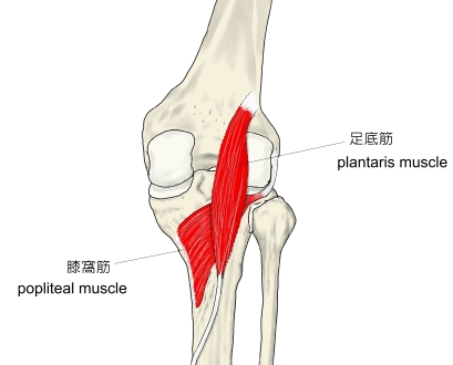 |
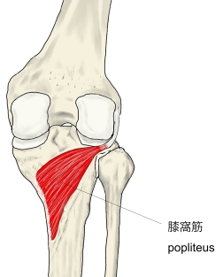
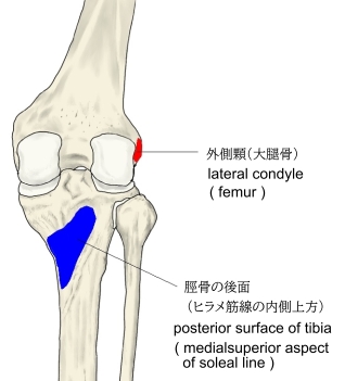
【 起 始 】: 大腿骨の外側顆、外側側副靭帯、膝関節包
【 停 止 】: 脛骨の後面(ヒラメ筋線の方外側より)
 |
「膝関節を屈曲し、下腿を内方に回転する。 」 ( 日本人体解剖学 )
・ 神 経 : 脛骨神経(L4,L5,S1)
 |
The popliteus muscle in the leg is used for unlocking the knees when walking, by laterally rotating the femur on the tibia during the closed chain portion of the gait cycle (one with the foot in contact with the ground). In open chain movements (when the involved limb is not in contact with the ground), the popliteus muscle medially rotates the tibia on the femur. It is also used when sitting down and standing up. It is the only muscle in the posterior (back) compartment of the lower leg that acts just on the knee and not on the ankle. The gastrocnemius muscle acts on both joints.
【Structure】
The popliteus muscle originates from the lateral surface of the lateral condyle of the femur by a rounded tendon. Its fibers pass downward and medially. It inserts onto the posterior surface of tibia, above the soleal line. The muscle arises within the capsule of knee joint and its tendon separates the lateral meniscus from the lateral ligament of the joint.
【Nerve supply】
The popliteus muscle is supplied by the tibial nerve, from spinal roots L5 and S1.
【Variation】
There is sometimes an additional head from the sesamoid bone in the lateral (outer) head of the gastrocnemius muscle.
Rarely an additional inconstant muscle; the popliteus minor is seen. It originates from the femur on the inner side of the plantaris muscle and inserts into the posterior ligament of the knee-joint.
Peroneotibialis, 14% of population. Origin is inner side of the head of the fibula, insertion into the upper end of the oblique line of the tibia, it lies beneath the popliteus.
Another variant, the cyamella, is a small sesamoid bone embedded in the tendon of the popliteus muscle. It is rarely seen in humans, but has been described more often in other primates and certain other animals. [3]
【Function】
The popliteus assists in flexing the leg upon the thigh; when the leg is flexed, it will rotate the tibia inward.
It is especially called into action at the beginning of the act of bending the knee, in as much as it produces the slight inward rotation of the tibia, which is essential in the early stage of this movement.
When the knee is in full extension, the femur slightly medially rotates on the tibia to lock the knee joint in place. Popliteus is often referred to as the "Key" to unlocking the knee since it begins knee flexion by laterally rotating the femur on the tibia.
Popliteus is also attached to the lateral meniscus in the knee and draws it posteriorly during knee flexion to prevent crushing the meniscus between the tibia and femur as the knee flexes.
【 語 句 】
・femur:大腿骨 ・tibia:脛骨 ・gait cycle:歩行周期 ・gastrocnemius muscle:腓腹筋 ・lateral condyle:外側顆 ・soleal line:ヒラメ筋線 ・capsule:関節包 ・meniscus:半月板 ・tibial nerve:脛骨神経 ・sesamoid bone:豆状骨 ・embed:はめ込む ・primate:霊長類
![]()