

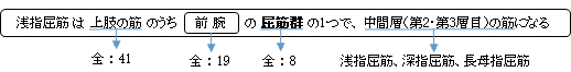
浅指屈筋 ( せんしくっきん、英:flexor digitorum superficialis muscle )
・ 概 要 |
・ 作 用 |
・ イラスト掲載サイト |
|
・ イラスト |
・ 神経 / 脈管 |
||
・ 起始 / 停止 |
・ Wikipedia |
![]()
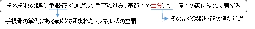
基節骨上で二分するときに(又はその先で)腱束の一部は交叉をする。 ⇒ 腱交叉 ( chiasm of Camper )
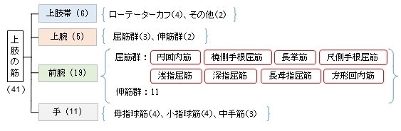
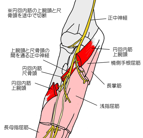
・ 筋腹の多くは第1層の筋(円回内筋、尺側手根屈筋、長掌筋、橈側手根屈筋)に覆われる形になる。
触察 : 握りこぶしを作り力いっぱい手首を手前に曲げたときに、外側から橈側手根屈筋 長掌筋、浅指屈筋
の腱と浮 き出るので、容易に確認ができる。(浮き出ない場合でも触診することにより確認可能)
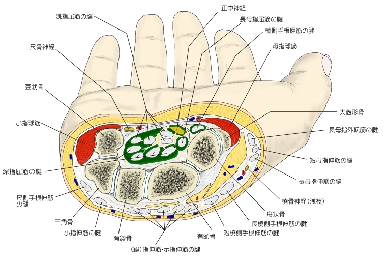
⇒ イラスト解説
筋連結 : 円回内筋、 上腕ニ頭筋、 尺側手根屈筋、 橈側手根屈筋、 長掌筋
「日本人体解剖学 (上巻) 」には以下のような解説が見られる。
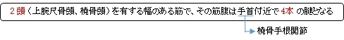
「2頭は合っして幅の広い筋腹を作り、橈側手根屈筋および長掌筋の下を下行して4本の腱に分かれる。さらに、第3、第4指への腱は長い層を、第2、第5指への腱は深い層を下行する。4腱はいずれも屈筋支帯の下にある手根管を通って手掌にいく。」
「異常/変異」ということで以下のような資料に解説が見られる。
「4個の独立した筋に分かれること、橈骨頭が小さくあるいは欠けること。第5指腱の欠けること。腱が手掌腱膜に連なること。深指屈筋から筋束を受けることなどがある。」「日本人体解剖学」(南山堂)
「橈側頭の強さはすこぶるまちまちである.これは全く欠如していることがある.示指にゆく筋腹はしばしば完全に独立している.Graeper(Anat. Anz., 50. Bd.,1917)は示指にいたる独立の筋腹を手掌でみている.ずっと下方に変位したこの筋腹の上方につづく腱はその上腕頭の内部で尺側上顆まで追跡された.下方の腱は通常の浅指屈筋の腱と同じ関係であった.4つの筋腹が全部独立していることはいっそうまれである.小指にゆく腱はしばしば欠けている.隣接する筋との結合は比較的しばしばみられる.この筋はまれに1つの腱を手掌腱膜に送っている.」(Rauber-Kopsch 解剖学)
![]()
 |
 |
||||
![]()
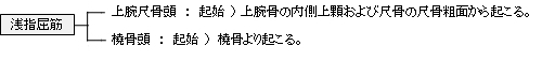
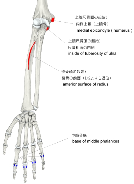
【 起 始 】 : ・上腕尺骨頭 :上腕骨の内側上顆、尺骨の粗面(内側)
・ 橈骨頭 : 橈骨の前面(1/2よりも近位)
【 停 止 】 : 第2~第5中節骨の骨底
 |
![]()
第2~第5指の第2関節(近位指節間関節)を屈曲させる。
※ Wikipediaでは上記の関節以外に 「中手指節関節」 や 「橈骨手根関節 (手首の関節)」 の名称も見られる。
![]()
・ 神 経 : 正中神経 (C7, C8, T1)
 |
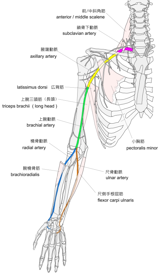 |
![]()
Flexor digitorum superficialis (flexor digitorum sublimis) is an extrinsic flexor muscle of the fingers at the proximal interphalangeal joints.
It is in the anterior compartment of the forearm. It is sometimes considered to be the deepest part of the superficial layer of this compartment, and sometimes considered to be a distinct, "intermediate layer" of this compartment. It is relatively common for the Flexor digitorum superficialis to be missing from the little finger, bilaterally and unilaterally, which can cause problems when diagnosing a little finger injury.
【 Structure 】
The muscle has two classically described heads – the humeroulnar and radial – and it is between these heads that the median nerve and ulnar artery pass. The ulnar collateral ligament of elbow joint gives its origin to part of this muscle.
Four long tendons come off this muscle near the wrist and travel through the carpal tunnel formed by the flexor retinaculum. These tendons, along with those of flexor digitorum profundus, are enclosed by a common flexor sheath. The tendons attach to the anterior margins on the bases of the intermediate phalanges of the four fingers. These tendons have a split (Camper's Chiasm) at the end of them through which the tendons of flexor digitorum profundus pass.
【 Innervation 】
The Flexor digitorium superficialis muscle is innervated by the median nerve (C7, C8, T1).
【 Function 】
The primary function of flexor digitorum superficialis is flexion of the middle phalanges of the four fingers (excluding the thumb) at the proximal interphalangeal joints, however under continued action it also flexes the metacarpophalangeal joints and wrist joint.
To test flexor digitorum superficialis, one finger is flexed at the proximal interphalangeal joint against resistance, while the remaining three fingers are held fully extended (to inactivate flexor digitorum profundus).
【 語 句 】
・ extrinsic : 外来的な ・ proximal interphalangeal joints : 近位指節間関節 ・ distinct : 全く別の、明瞭な ・ relatively : 相対的に ・ bilaterally : 両側的に ・ unilaterally : 一側性に ・ diagnose : 診断する ・ humeroulnar : 上腕尺骨(頭) ・ radial : 橈骨(頭) ・ median nerve : 正中神経 ・ ulnar artery : 尺骨動脈 ・ ulnar collateral ligament : 内側側副靭帯 ・ carpal tunnel : 手根管 ・ flexor retinaculum : 屈筋支帯 ・ flexor digitorum profundus : 深指屈筋 ・ sheath : 鞘 ・ intermediate phalanges : 中節骨 ・ middle phalanges : 中節骨 ・ proximal interphalangeal joints : 近位指節間関節 ・ metacarpophalangeal joints : 中手指節関節
![]()
![]()