

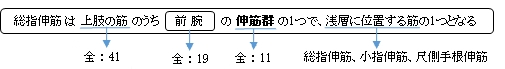
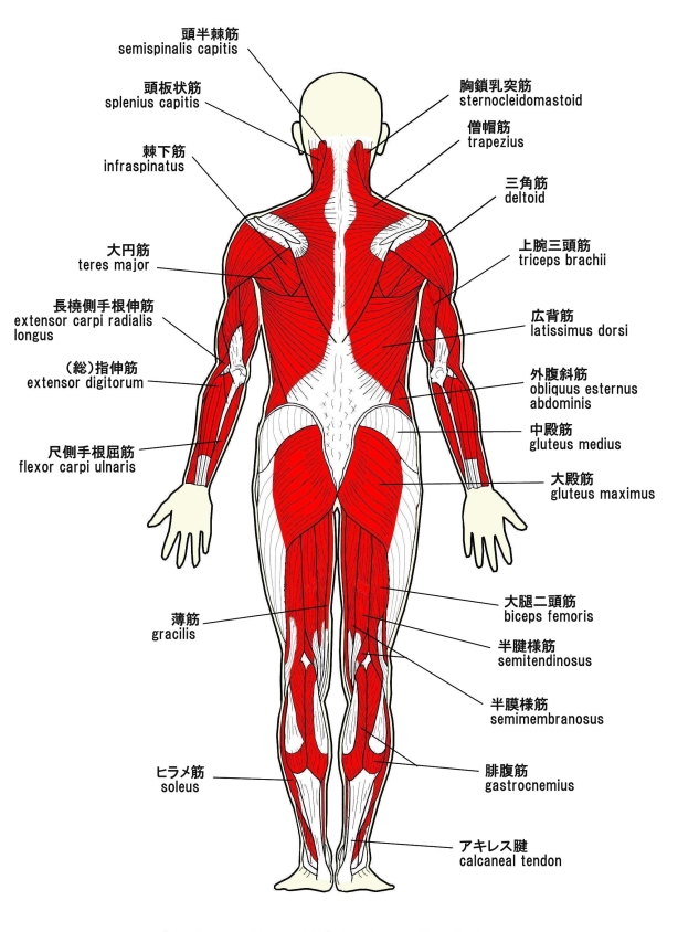
(総)指伸筋 ( そうししんきん、英:extensor digitorum muscle )
・ 概 要 |
・ 作 用 |
・ イラスト掲載サイト |
|
・ イラスト |
・ 神経 / 脈管 |
||
・ 起始 / 停止 |
・ Wikipedia |
![]()
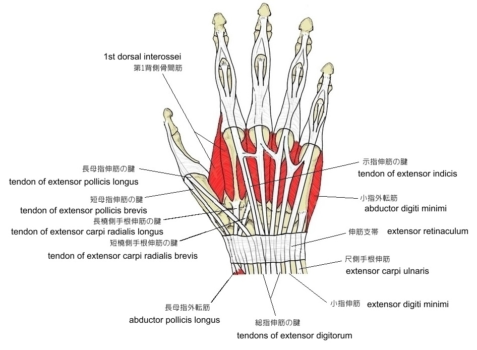
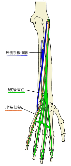
触察 : 腱は手背において容易に確認ができ、また、筋腹も、皮下において2つほど確認が可能となる。
⇒ 腱のイラスト解説
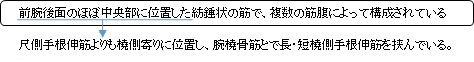
筋連結 : 尺側手根伸筋、 示指伸筋、 回外筋、 短橈側手根伸筋、 小指伸筋
「Rauber-Kopsch解剖学」には以下のような解説が見られる。
「 変異 : その腱についてはすでに述べた事がらのほかに,1つの過剰の腱が母指に達していることがある.(日本人における総指伸筋の第4腱の欠如は男208体側のうち17体側(8.2%),女93体側のうち15体側(16.1%)である(小金井良精・新井春次郎,敷波重次郎:東京医学会雑誌,17巻,127~131,1903).)」
![]()
 |
 |
 |
|||
 |
 |
||||
![]()
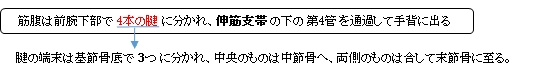
備考 : 腱の末端は基節骨底で3つに分かれ、 中央のものが中節骨骨底で、その両側 のものは末節骨骨底で停止する。
 |
![]()
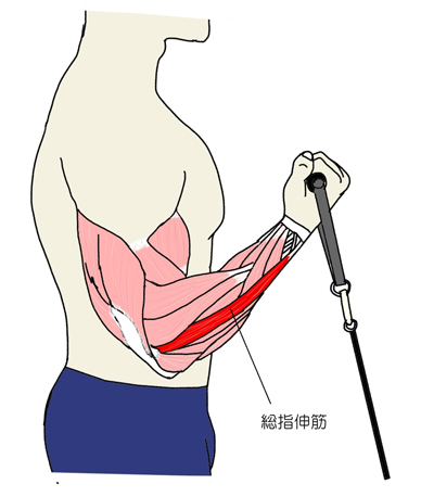
「 第2~第5指を伸ばし、同時に手根も伸ばす。 」 ( 日本人体解剖学 )
![]()
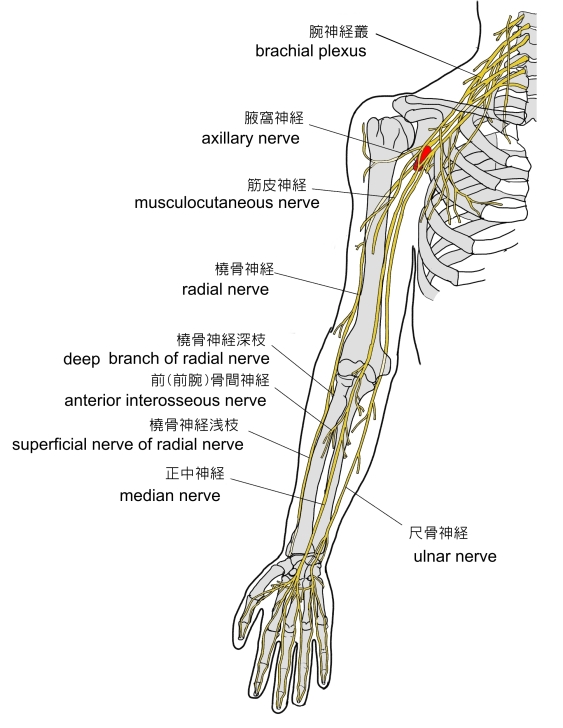
・ 神 経 : 橈骨神経(深枝) (C6~C8)
・ 脈 管 : 後骨間動脈
 |
 |
![]()
The extensor digitorum muscle (also known as extensor digitorum communis) is a muscle of the posterior forearm present in humans and other animals. It extends the medial four digits of the hand. Extensor digitorum is innervated by the posterior interosseous nerve, which is a branch of the radial nerve.
【 Structure 】
The extensor digitorum muscle arises from the lateral epicondyle of the humerus, by the common tendon; from the intermuscular septa between it and the adjacent muscles, and from the antebrachial fascia. It divides below into four tendons, which pass, together with that of the extensor indicis proprius, through a separate compartment of the dorsal carpal ligament, within a mucous sheath. The tendons then diverge on the back of the hand, and are inserted into the middle and distal phalanges of the fingers in the following manner.
Opposite the metacarpophalangeal articulation each tendon is bound by fasciculi to the collateral ligaments and serves as the dorsal ligament of this joint; after having crossed the joint, it spreads out into a broad aponeurosis, which covers the dorsal surface of the first phalanx and is reinforced, in this situation, by the tendons of the interossei and lumbricalis.
Opposite the first interphalangeal joint this aponeurosis divides into three slips; an intermediate and two collateral: the former is inserted into the base of the second phalanx; and the two collateral, which are continued onward along the sides of the second phalanx, unite by their contiguous margins, and are inserted into the dorsal surface of the last phalanx. As the tendons cross the interphalangeal joints, they furnish them with dorsal ligaments. The tendon to the index finger is accompanied by the tendon of extensor indicis, which lies on its ulnar side. On the back of the hand, the tendons to the middle, ring, and little fingers are connected by two obliquely placed bands, one from the third tendon passing inferior and laterally to the second tendon, and the other passing from the same tendon inferior and medially to the fourth.
The extensor tendons are connected to the second by a thin transverse band, known as the juncturae tendinum; they serve to maintain the central alignment of the extensor tendons over the metacarpal head, thus increasing the available leverage. Injuries (such as by an external flexion force during active extension) may allow the tendon to dislocate into the intermetacarpal space; the extensor tendon then acts as a flexor and the finger may no longer be actively extended. This may be corrected surgically by using a slip of the extensor tendon to replace the damaged ligamentous band.
【 Function 】
The extensor digitorum muscle extends the phalanges, then the wrist, and finally the elbow. It tends to separate the fingers as it extends them.
In the fingers, the extensor digitorum acts principally on the proximal phalanges, acting to extend the metacarpophalangeal joint. Extension of the proximal and distal interphalangeal joints, however, is mediated predominantly by the dorsal and palmar interossei and lumbricals of the hand.
【 語 句 】
・ digit : 指 ・ posterior interosseous nerve : 後骨間神経 ・ radial nerve : 橈骨神経 ・ lateral epicondyle : 外側上顆 ・ humerus : 上腕骨 ・ intermuscular septa : 筋間中隔 ・ adjacent : 近隣の ・ antebrachial fascia : 前腕筋膜 ・ extensor indicis proprius : 示指伸筋 ・ dorsal carpal ligament : 背側手根靭帯? ・ mucous : 粘液を分泌する ・ sheath : 鞘 ・ distal phalanges : 末節骨 ・ in the following manner : 以下のやり方で ・ metacarpophalangeal : 中手指節関節の ・ articulation : 関節(接合) ・ fasciculi : 束 ・ aponeurosis : 腱膜 ・ phalanx : 篩骨 ・ reinforce : 補強する ・ interossei : interosseus(骨間筋)の複数形 ・ lumbricalis : 虫様筋 ・ interphalangeal joint : 指節間関節 ・ contiguous : 接触する、連続した ・ furnish : 供給する、取り付ける ・ index finger : 人差し指 ・ extensor indicis : 示指伸筋 ・ ulnar : 尺骨の ・ juncturae tendinum : 県間結合 ・ leverage : てこの力 ・ dislocate : 脱臼させる ・ proximal phalanges : 末節骨 ・ mediated : 取り次ぐ ・predominantly : 主に ・ palmar interossei : 掌側骨間筋
![]()
![]()