

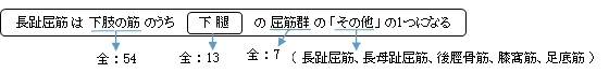
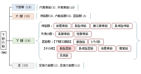
長趾屈筋 ( ちょうしくっきん、英:flexor digitorum longus muscle )
・ 概 要 |
・ 作 用 |
・ イラスト掲載サイ |
|
・ イラスト |
・ 神経 / 脈管 |
||
・ 起始 / 停止 |
・ Wikipedia |
 。
。
以下は「船戸和也のHP」の解説文となる。
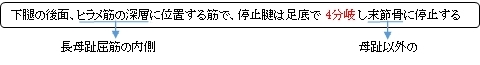
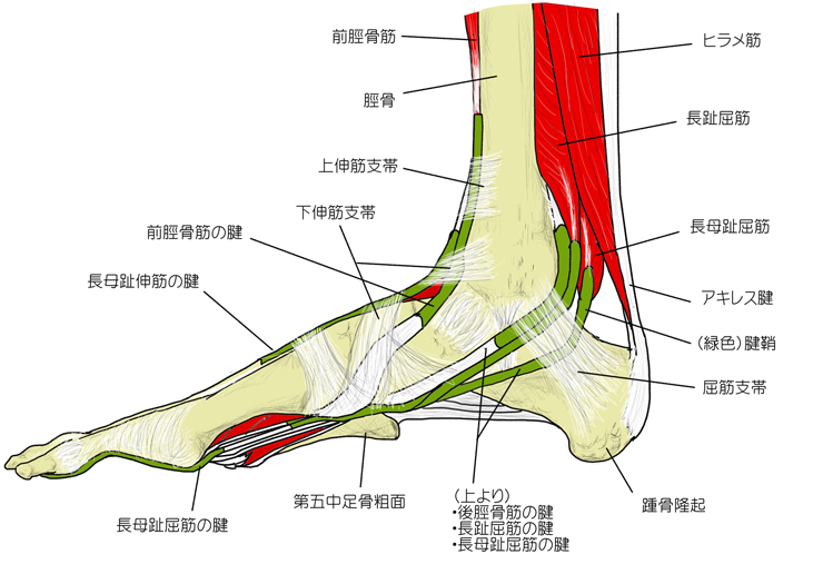
「長趾屈筋はヒラメ筋線より遠位の脛骨後面およびヒラメ筋腱弓の一部から起こる。その停止腱は基節骨の領域で短趾屈筋の腱を貫通し(第2~5)趾の末節骨に停止する。長趾屈筋の腱は腱鞘に包まれて、内果溝を後脛骨筋腱の背外側に走り、屈筋死体の下を載距突起内側縁に沿って足底に至る。舟状骨粗面のレベルでは長母趾屈筋腱の浅層を通る。この際、長母趾屈筋の健束が長趾屈筋の腱に混じる。この腱交叉位遠では足底方形筋が長趾屈筋の腱に付く。この付加的な屈筋は長趾屈筋停止腱の牽引方向を趾放線の長軸方向と関連させる。同一趾へ向かう長趾屈筋(「貫通筋」と短趾屈筋「被貫通筋」)の停止腱は腱鞘(滑液鞘に包まれる。腱鞘は第1中足骨頭の上方から始まり、末節骨までのびている。これらの滑液鞘は線維鞘に包まれる。線維鞘は手指におけると同じように横走線維と交織する線維(輪状および十字部)からなる。」
 |
 |
 |
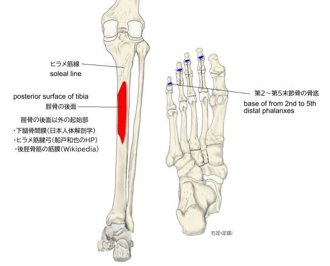
【 起 始 】: 脛骨の後面(ヒラメ筋線より遠位)、下腿骨間膜
※「船戸和也のHP」では、起始部には下腿骨間膜の名称は見られず「ヒラメ筋腱弓の一部」も起始の一つとしている。
※Wikipediaでは起始部を以下のように解説している。
・ヒラメ筋線の下端よりも7~8㎝以内の脛骨の後面(後脛骨筋の起始部よりも内側)
・後脛骨筋の筋膜
【 停 止 】: (短趾屈筋の腱を貫通して)第2~第5末節骨の骨底
⇒ 長趾屈筋の腱が短趾屈筋の腱を貫通しているのがわかるイラスト
 |
「 第2~第5指を屈曲し、足を足底側に曲げる。」 ( 日本人体解剖学 )
・ 神 経 : 脛骨神経(L5,S1,S2) ※資料によっては(L4~S1)
・ 脈 管 : 後脛骨動脈
The flexor digitorum longus muscle is situated on the tibial side of the leg. At its origin it is thin and pointed, but it gradually increases in size as it descends. It serves to flex the second, third, fourth, and fifth toes.
【Structur】
The flexor digitorum longus muscle arises from the posterior surface of the body of the tibia, from immediately below the soleal line to within 7 or 8 cm of its lower extremity, medial to the tibial origin of the tibialis posterior muscle. It also arises from the fascia covering the tibialis posterior muscle.
The fibers end in a tendon, which runs nearly the whole length of the posterior surface of the muscle. This tendon passes behind the medial malleolus, in a groove, common to it and the tibialis posterior, but separated from the latter by a fibrous septum, each tendon being contained in a special compartment lined by a separate mucous sheath. The tendon of the tibialis posterior and the tendon of the flexor digitorum longus cross each other, in a spot above the medial malleolus, the crural tendinous chiasm. It passes through the tarsal tunnel.
It passes obliquely forward and lateralward, superficial to the deltoid ligament of the ankle-joint, into the sole of the foot, where it crosses over the tendon of the flexor hallucis longus at the level of the navicular bone at a location known as the knot of henry (also referred to as plantar tendinous chiasm), and receives from it a strong tendinous slip.
It then expands and is joined by the quadratus plantæ muscle, and finally divides into four tendons, which are inserted into the bases of the last phalanges of the second, third, fourth, and fifth toes, each tendon passing through an opening in the corresponding tendon of the flexor digitorum brevis muscle opposite the base of the first interphalangeal joint.
【 語 句 】
・tibial:脛骨の ・soleal line:ヒラメ筋線 ・extremity:末端 ・tibialis posterior muscle:後脛骨筋 ・fascia:筋膜 ・medial malleolus:内果 ・flexor digitorum longus:長趾伸筋 ・chiasm:交叉 ・tarsal tunnel:足根管 ・deltoid ligament:三角靭帯 ・flexor hallucis longus:長母趾屈筋 ・navicular bone:舟状骨 ・quadratus plantæ muscle:足底方形筋 ・phalanges:趾節骨 ・flexor digitorum brevis muscle:短趾屈筋 ・interphalangeal joint:趾節間関節
【Variation】
Flexor accessorius longus digitorum, not infrequent, origin from fibula, or tibia, or the deep fascia and ending in a tendon which, after passing beneath the laciniate ligament, joins the tendon of the long flexor or the quadratus plantæ.
【Function】
Similar to the flexor hallucis longus and tibialis posterior muscles, the flexor digitorum longus muscle functions to plantar flex and invert the foot. The flexor digitorum longus muscle is responsible for the movement and curling of the second, third, fourth and fifth toes. This muscle makes it possible for the toes to grip the surface of floors, which is important when it comes to maintaining postural balance on surfaces that are rough or uneven. The other deep muscles are the flexor hallucis longus and tibialis posterior; the tibialis posterior is the most powerful of these deep muscles. All three muscles are innervated by the tibial nerve which comprises half of the sciatic nerve.
【 語 句 】
・fibula:腓骨 ・laciniate:条裂? ・tibial nerve:脛骨神経 ・comprise:構成する ・sciatic nerve:坐骨神経
![]()