

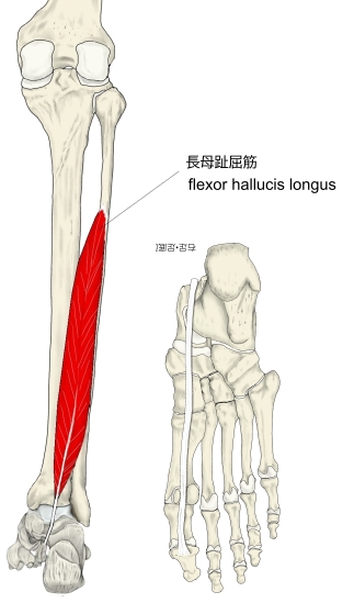
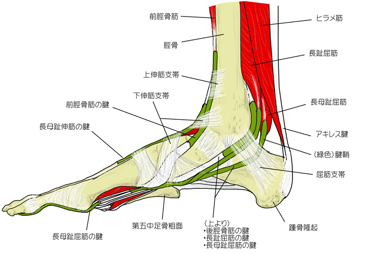
長母趾屈筋 ( ちょうぼしくっきん、英:flexor hallucis longus muscle )
・ 概 要 |
・ 作 用 |
・ イラスト掲載サイ |
|
・ イラスト |
・ 神経 / 脈管 |
||
・ 起始 / 停止 |
・ Wikipedia |
・「長母趾屈筋の筋腹は、アキレス腱の深層で踵骨の近位縁から約1横指頭方の部位まで存在する。」(骨格筋の形と触察法)
筋連結 : 長趾屈筋、後脛骨筋、長腓骨筋、短腓骨筋、前脛骨筋、長母趾伸筋、
以下は「船戸和也のHP」の解説文となる。
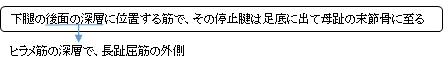
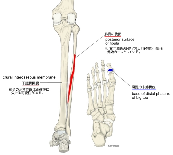
「長母趾屈筋はずっと内側に(おもな腱は第1末節骨底に)停止する。その起始は下腿の外側、つまり腓骨後面の遠位2/3,骨間膜の狭い紐状部分および後筋間中隔である。その腱は長趾屈筋腱の下を横切り(足底腱交叉)、足底で第2および第3(まれに第4)趾末節骨に腱性停止を送る。距骨後面と載距突起下面において長母趾屈筋の腱は溝の中を走る。同筋は腱鞘に包まれる。腱鞘は内果先端のレベルにはじまり、遠位へ伸び、第1中足骨底に至る。停止腱は第1中足骨頭から末節骨に至るまで腱鞘に包まれている。腱鞘は線維性のおおいによって母趾の各分節に付く。」
 |
 |
 |
※「船戸和也のHP」では上記の2つのほかに「後筋間中隔」を加えている。
※「腓骨の後面」に関してWikipediaには以下のような解説文が見られる。
「It arises from the inferior two-thirds of the posterior surface of the body of the fibula」
【 停 止 】: 母指の末節骨、骨底
 |
「 母指を足底側に屈曲する。」 ( 日本人体解剖学 )
・ 神 経 : 脛骨神経(L5,S1,S2)
・ 脈 管 : 腓骨動脈
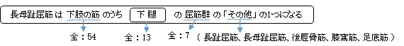
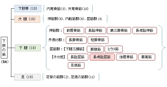
The flexor hallucis longus muscle (FHL) is one of the three deep muscles of the posterior compartment of the leg that attaches to the plantar surface of the distal phalanx of the great toe. The other deep muscles are the flexor digitorum longus and tibialis posterior; the tibialis posterior is the most powerful of these deep muscles. All three muscles are innervated by the tibial nerve which comprises half of the sciatic nerve.
【Structure】
The flexor hallucis longus is situated on the fibular side of the leg. It arises from the inferior two-thirds of the posterior surface of the body of the fibula, with the exception of 2.5 cm. at its lowest part; from the lower part of the interosseous membrane; from an intermuscular septum between it and the peronius muscles, laterally, and from the fascia covering the tibialis posterior, medially.
The fibers pass obliquely downward and backward, where it passes through the tarsal tunnel on the medial side of the foot and end in a tendon which occupies nearly the whole length of the posterior surface of the muscle.
This tendon lies in a groove which crosses the posterior surface of the lower end of the tibia, between the medial and lateral tubercles of the posterior surface of the talus, and the under surface of the sustentaculum tali of the calcaneus; in the sole of the foot it runs forward between the two heads of the flexor hallucis brevis, and is inserted into the base of the last phalanx of the great toe. The grooves on the talus and calcaneus, which contain the tendon of the muscle, are converted by tendinous fibers into distinct canals, lined by a mucous sheath.
As the tendon passes forward in the sole of the foot, it is situated above, and crosses from the lateral to the medial side of the tendon of the flexor digitorum longus, to which it is connected by a fibrous slip.
【Variation】
Usually a slip runs to the flexor digitorum and frequently an additional slip runs from the flexor digitorum to the flexor hallucis. Peroneocalcaneus internus, rare,[clarification needed] arises below or outside the flexor hallucis from the back of the fibula, passes over the sustentaculum tali with the flexor hallucis and inserts into the calcaneum.
【Function】
Similar to the flexor digitorum longus and tibialis posterior muscles, the flexor hallucis longus muscle functions to plantar flex and invert the foot. However, it is unique in that it also functions to flex the great toe and helps supinate the ankle.
【 語 句 】
・distal phalanx:末節骨 ・flexor digitorum longus:長趾屈筋 ・tibialis posterior:後脛骨筋 ・tibial nerve:脛骨動脈 ・comprises:構成する ・sciatic nerve:坐骨神経 ・fibula:腓骨 ・interosseous membrane:骨間膜 ・intermuscular septum:筋間中隔 ・peronius muscles:腓骨筋群 ・fascia:筋膜 ・tarsal tunnel:足根管 ・groove:溝 ・tubercle:結節 ・talus:距骨 ・sustentaculum tali:載距突起 ・calcaneus:踵骨 ・flexor hallucis brevis:短母趾屈筋 ・phalanx:指骨 ・distinct:全く別の、はっきりした ・mucous:粘液を分泌する ・calcaneum:踵骨 ・supinate:回外させる
![]()