

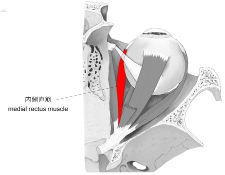
内側直筋 ( ないそくちょくきん、 英 : medial rectus muscle )
・ 概 要 |
・ 作 用 |
・ イラスト掲載サイ |
|
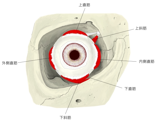
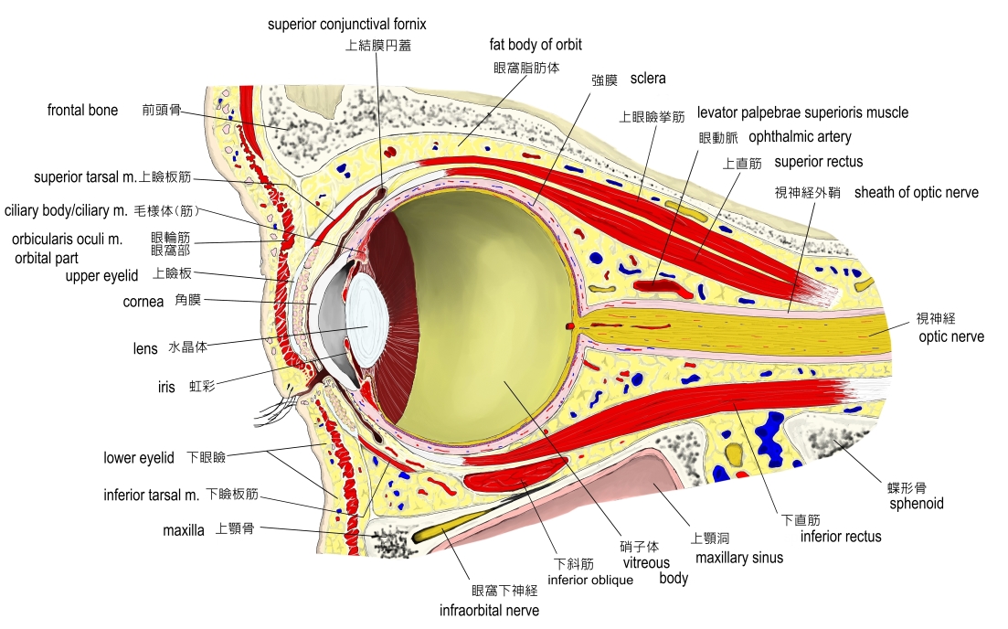
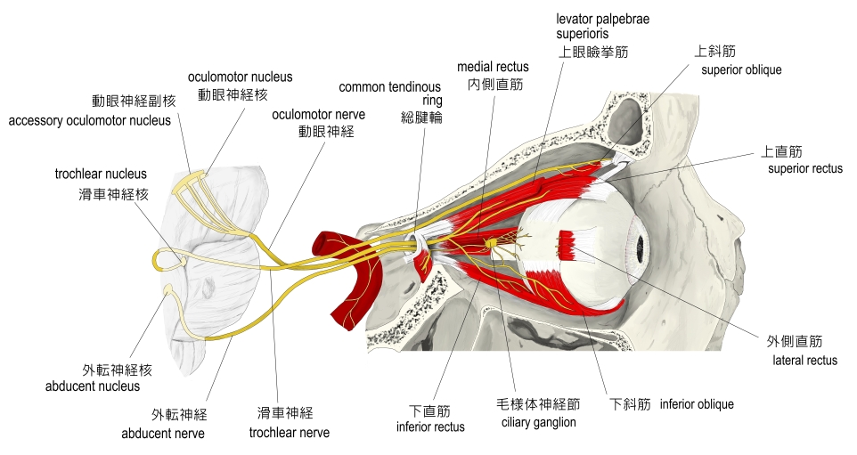
・ イラスト |
・ 神経 / 脈管 |
||
・ 起始 / 停止 |
・ Wikipedia |

・ 「 眼筋のうち最強のもので、長さは上直筋に次ぐが、腱は最も短く、眼窩の内側壁に沿って走る。 」 ( 日本人体解剖学 )
・ いわゆる「 眼 筋 」の1つになる。
 |
|
|
|
|
【 起 始 】: 「 総腱輪から起こり、眼窩の内側壁に沿って前方に進む。」 ( 日本人体解剖学 )
総腱輪( anulus tendineus ) : 眼窩の後方にある視神経を輪状に囲んでいる4つの直筋群の起始部となる部分
【 停 止 】: 「 強膜内側方の赤道前部 」 ( 日本人体解剖学 )
「 眼球の前極を内側方(内転)に引く。」 ( 日本人体解剖学 )
・神 経 : 動眼神経 (第3脳神経)
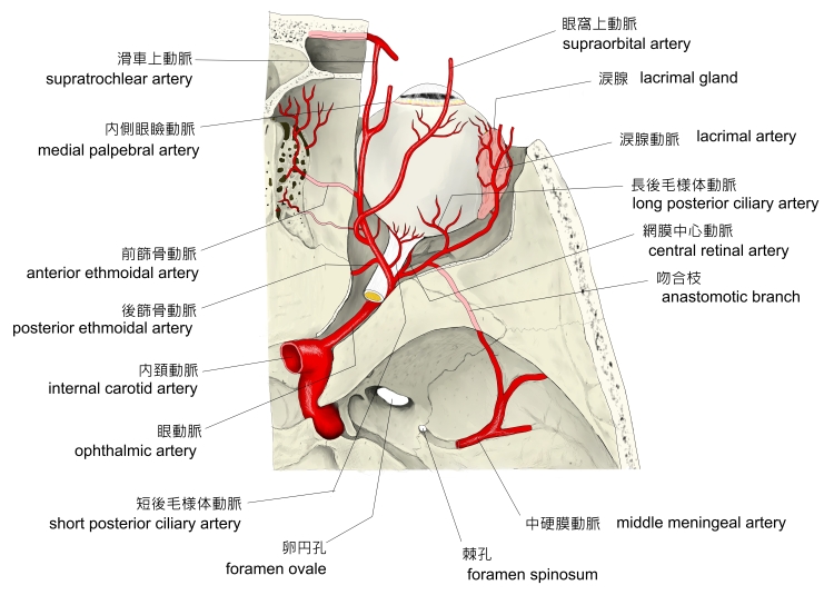
・脈 管 : 動脈)前毛様体動脈(眼動脈の枝)? 恐らく静脈もそれに準じるものだと思われる。
以下は「Wikipedia」の「anterior ciliary arteries(前毛様体動脈)」の解説文の一部となる。
「Three of the four rectus muscles; the superior, inferior and medial, are supplied by two ciliary
arteries each, while the lateral rectus only receives one branch.」
|
 |
|
||
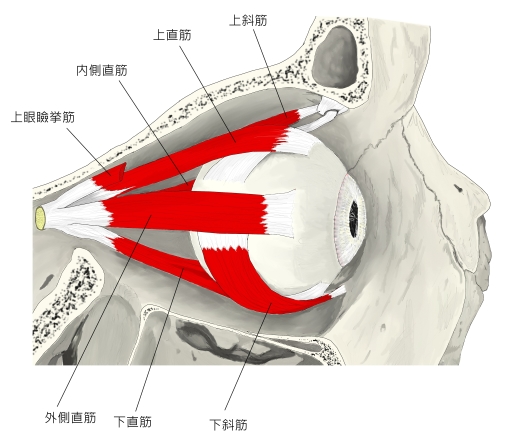
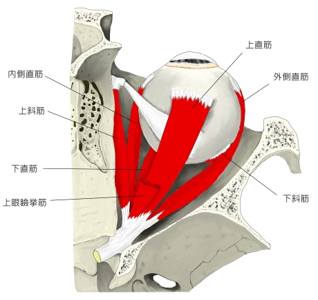
右眼窩(外側面より) |
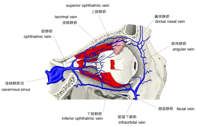
右眼窩(上面より) | 右眼窩の静脈 |
The medial rectus muscle is a muscle in the orbit.
As with most of the muscles of the orbit, it is innervated by the inferior division of the oculomotor nerve ( Cranial Nerve III ).
This muscle shares an origin with several other extrinsic eye muscles, the anulus tendineus, or common tendon.
It is shorter but stronger than the other orbital recti muscles.[1]
【 語 句 】
・ orbit : 眼窩 ・ oculomotor nerve : 動眼神経 ・ Cranial Nerve : 脳神経 ・ extrinsic : 外部の ・ anulus tendineus : 総腱輪 ・ recti muscles : 直筋
![]()