

上斜筋 ( じょうしゃきん、 英 : superior oblique muscle )
・ 概 要 |
・ 作 用 |
・ イラスト掲載サイ |
|
・ イラスト |
・ 神経 / 脈管 |
||
・ 起始 / 停止 |
・ Wikipedia |
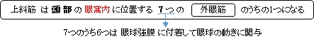
・ 「 眼筋のうち腱も含めれば最も長い筋である。 」 ( 日本人体解剖学 )
・ いわゆる「 眼 筋 」の1つになる。
 |
 |
 |
|
|
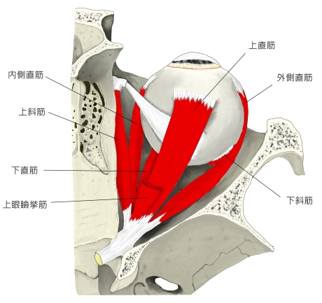
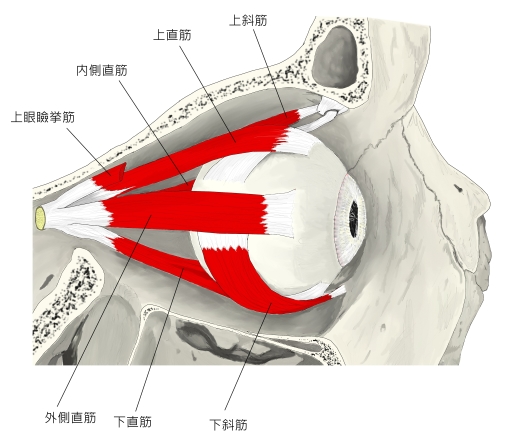
【 起 始 】: 「 総腱輪から起こり、眼窩の内側壁と上壁との境に沿い上直筋・内側直筋の上を前進し、前頭骨滑車窩にあって繊維軟骨輪である滑車内を通る。この部位にある粘液嚢を滑車滑液包という。滑車を出ると、上斜筋の腱は眼球に向かい次第に幅が広く、かつ薄くなって斜め後外側方に走り、上直筋の下をこれと交差するように進む。」 ( 日本人体解剖学 )
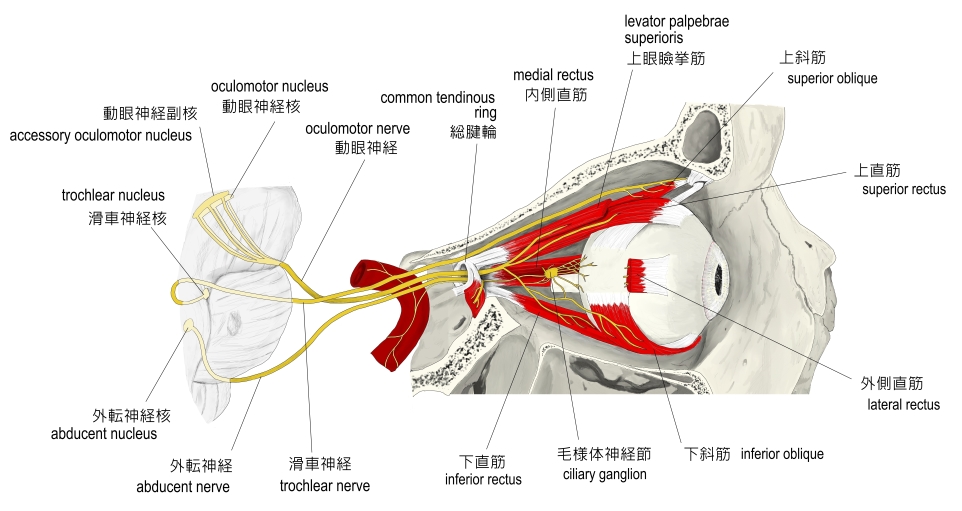
総腱輪( anulus tendineus ) : 眼窩の後方にある視神経を輪状に囲んでいる4つの直筋群の起始部となる部分
【 停 止 】: 「 強膜上面の赤道後部 」 ( 日本人体解剖学 )
「 眼球の前極を下外側方に(下転および外転)、上極を内側方に引く。(内側方回転) 」 ( 日本人体解剖学 )
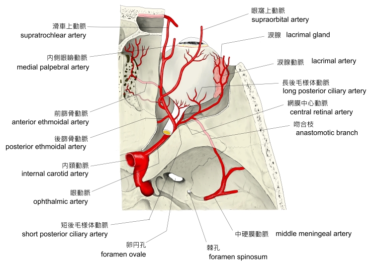
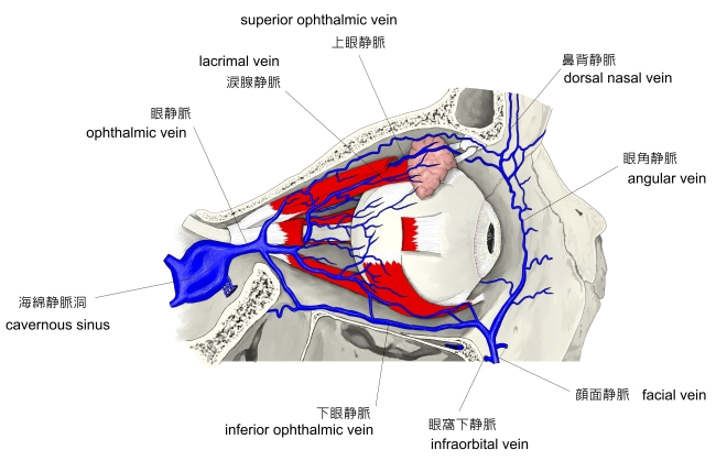
・ 脈 管 : 上の神経より、おそらく同名の 眼動脈 および 眼静脈(の枝?)だと思われるが、それらに言及している資料は見当たらない。
|
 |
|
The superior oblique muscle, or obliquus oculi superior, is a fusiform muscle originating in the upper, medial side of the orbit (i.e. from beside the nose) which abducts, depresses and internally rotates the eye. It is the only extraocular muscle innervated by the trochlear nerve ( the fourth cranial nerve ).
See also: Extraocular muscles
The superior oblique muscle loops through a pulley-like structure ( the trochlea of superior oblique) and inserts into the sclera on the posterotemporal surface of the eyeball. It is the pulley system that gives superior oblique its actions, causing depression of the eyeball despite being inserted on the superior surface.
The superior oblique arises immediately above the margin of the optic foramen, superior and medial to the origin of the superior rectus, and, passing forward, ends in a rounded tendon, which plays in a fibrocartilaginous ring or pulley attached to the trochlear fossa of the frontal bone.
The contiguous surfaces of the tendon and ring are lined by a delicate mucous sheath, and enclosed in a thin fibrous investment.
The tendon is reflected caudally, laterally, and inferiorly beneath the superior rectus to the lateral part of the bulb of the eye, and is inserted onto the scleral surface, behind the equator of the eyeball, the insertion of the muscle lying between the superior rectus and lateral rectus.
【 語 句 】
・ fusiform muscle:紡錘状筋 ・ orbit: ・ abduct: ・extraocular muscle : ・trochlear nerve : ・ cranial nerve: ・ pulley:滑車 ・ trochlea:滑車 ・ sclera:強膜 ・ optic foramen: 視神経孔 ・ fibrocartilaginous:線維軟骨の ・ trochlear fossa:滑車窩 ・contiguous :隣接する ・investment :外皮、外層 ・ caudally:尾側に ・ equator:赤道
See also: Eye movement
The primary ( main ) action of the superior oblique muscle is intorsion (internal rotation),[1] the secondary action is depression ( primarily in the adducted position ) and the tertiary action is abduction (lateral rotation).
The extraocular muscles rotate the eyeball around vertical, horizontal and antero-posterior axes. Extraocular muscles other than the medial rectus and lateral rectus have more than one action due to the angle they make with the optical axis of the eye while inserting into the eyeball. The superior and inferior oblique muscles make an angle of 51 degrees with the optical axis.[citation needed]
The depressing action of superior oblique (making the eye look down towards the mouth) is most effective when the eye is in an adducted position. This is because as the eye is abducted (looks laterally), the contribution made by superior oblique to depression of the eye decreases, as the inferior rectus muscle causes this movement more directly and powerfully. The main muscle for abduction is the lateral rectus, so although superior oblique contributes to a downwards and lateral eye movement, testing this motion would not be specific enough as inferior and lateral recti muscles would also be tested. Therefore, during neurological examinations, the superior oblique is tested by having the patient look inwards and downwards, testing only the depressing action of the muscle. This is a source of confusion on the subject as although clinical testing asks the patient to adduct and depress the eye, anatomically the muscle depresses and abducts it.
【 語 句 】
・ intorsion:内旋 ・adducted :内転させられた ・ tertiary:第3の ・ abduction:外転 ・ optical axis:光軸 ・ contribution:寄与 ・ neurological:神経学的な
The great importance of intorsion and extorsion produced by the two oblique muscles can only be understood when it is considered with regards to the other muscle actions present. The two obliques prevent the eye from rotating about its long axis ( retina to pupil ) when the superior and inferior rectus muscles contract. This is because the orbit does not face directly forwards- the centre-line of the orbit is a little over 20 degrees out from the mid-line.
But because the eyes do face forwards, when acting alone, as well as making the eye look up, superior rectus causes it to rotate slightly about the long axis, so the top of the eye moves medially (intorsion). Similarly, in addition to making the eye look down, inferior rectus would cause the eye to rotate about the long axis so the top of the eye moves slightly laterally (extorsion), if acting alone. Clearly this is undesirable as our vision would rotate when we looked up and down. For this reason, these two rectus muscles work in conjunction with the two obliques. When acting alone, superior oblique causes intorsion, inferior oblique, extorsion. Hence, when inferior rectus contracts so we look down, superior oblique also contracts to prevent extorsion of the eye, and when superior rectus contracts so we look up, inferior oblique contracts to prevent intorsion, thus the undesired rotatory actions of the inferior and superior recti about the long axis of the eye are cancelled out. This keeps our vision horizontally level, irrespective of eye position in the orbit.[2]
【 語 句 】
・ retina : 網膜 ・ contract : 収縮する ・ in conjunction with ~ : ~ と併せて ・ irrespective : 無関係の
![]()