

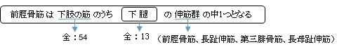
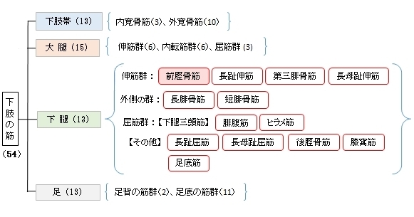
前脛骨筋 ( ぜんけいこつきん、英:tibialis anterior muscle )
・ 概 要 |
・ 作 用 |
・ イラスト掲載サイト |
|
・ イラスト |
・ 神経 / 脈管 |
||
・ 起始 / 停止 |
・ Wikipedia |
![]()
![]()
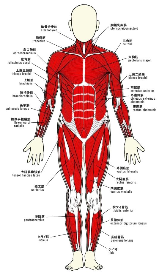
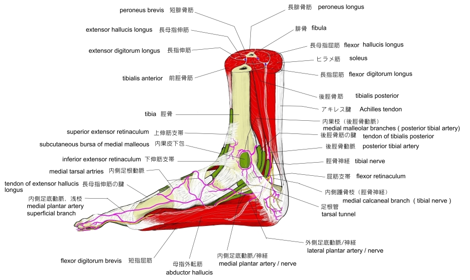
・いわゆるスネの横にある、足首を曲げたときにそのふくらみがしっかりと確認できる筋で、歩いたり走ったりするときによく使われる。
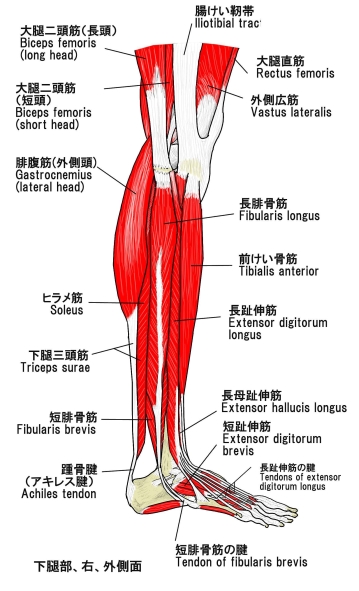
触察 : 脛骨のすぐ外側にあり、足首を上下に屈曲させることによってその筋腹が盛り上がるので、皮下において容易に触診することができる。
以下は「船戸和也のHP」の解説文となる。
「前脛骨筋は脛骨外側顆、脛骨外側面(近位2/3)、下腿筋膜および筋間膜から起始する。第1中足骨と第1楔状骨あたりの足底部に停止する。収縮中に筋腹は脛骨近位1/3の骨縁上に突出する。その腱は脛骨遠位1/3にかけて形成され、伸筋支帯の下を通って足の内側縁へ至る。その腱鞘は伸筋支帯より近位に始まり、距腿関節の関節腔のレベルにまで伸びている。腱鞘は前脛骨筋腱の遠位部および近位部浅層をおおい、中間部を包んでいる。前脛骨筋と長趾伸筋に対する近位の筋枝は深腓骨神経から同神経がまだ腓骨筋群を容れる部位を通っている内に分かれる。深腓骨神経が長趾伸筋を貫通してから遠位の筋枝が両筋の各々に行き(通常2条の)筋枝が母趾の伸筋へ行く。」
![]()
 |
 |
 |
|||
 |
|||||
![]()
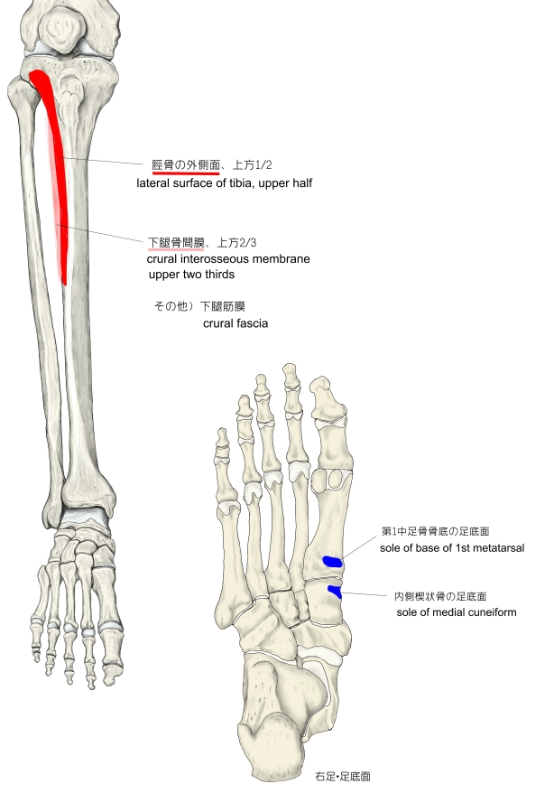
【 起 始 】 : 脛骨の外側面(上方1/2)、下腿骨間膜(上方2/3)、下腿筋膜
【 停 止 】 : 内側楔状骨の足底面、 第1中足骨の骨底の足底面
 |
![]()
「 足の内側縁をあげ(内反)、同時に足を足背面に屈曲する。足を固定すると、下腿を前方に傾ける。」 ( 日本人体解剖学 )
![]()
・ 神 経 : 深腓骨神経(L4,L5,S1)
・ 脈 管 : 前脛骨動脈、前脛骨反回動脈(前脛骨動脈の上方枝)
![]()
The tibialis anterior muscle is a muscle in humans that originates along the upper two-thirds of the lateral (outside) surface of the tibia and inserts into the medial cuneiform and first metatarsal bones of the foot. It acts to dorsiflex and invert the foot. This muscle is mostly located near the shin.
It is situated on the lateral side of the tibia; it is thick and fleshy above, tendinous below. The tibialis anterior overlaps the anterior tibial vessels and deep peroneal nerve in the upper part of the leg.
【Structure】
The tibialis anterior muscle arises from:
- the lateral condyle of the tibia.
- the upper 2/3 of the lateral surface of the tibia.
- the adjoining part of the interosseous membrane.
- the deep surface of the fascia.
- the intermuscular septum between it and the extensor digitorum longus.
The fibers of this circumpennate muscle are relatively parallel to the plane of insertion, ending in a tendon, apparent on the anteriomedial dorsal aspect of the foot close to the ankle.
It passes through the most medial compartments of the transverse and cruciate crural ligaments. It is inserted into the medial and under surface of the medial cuneiform bone and the base of the first metatarsal bone.
【 語 句 】
・tibia:脛骨 ・medial cuneiform bone:内側楔状骨 ・metatarsal bones:中足骨 ・fleshy:多肉質の ・peroneal nerve:腓骨神経 ・lateral condyle:外側顆 ・adjoining:隣の、付近の ・interosseous membrane:骨間膜 ・fascia:筋膜 ・intermuscular septum:筋間中隔 ・extensor digitorum longus:長趾伸筋 ・cruciate crural ligaments:?
【Nerve supply】
The tibialis anterior muscle is supplied by the deep fibular nerve , a branch of common fibular nerve.
【Variation】
A deep portion of the muscle is rarely inserted into the talus, or a tendinous slip may pass to the head of the first metatarsal bone or the base of the first phalanx of the great toe.
The tibiofascialis anterior, a small muscle from the lower part of the tibia to the transverse or cruciate crural ligaments or deep fascia.[clarification needed]
【Function】
The tibialis anterior muscle is the most medial muscle of the anterior compartment of the leg. It is responsible for dorsiflexing and inverting the foot, and is the largest dorsiflexor of the foot. The muscle has two origins, one being the lateral tibial condyle and the other being the upper lateral surface of the tibia, and inserts on the medial surface of the medial cuneiform and adjoining part of base of the first metatarsal of the foot allowing the toe to be pulled up and held in a locked position. It also allows for the ankle to be inverted giving the ankle horizontal movement allowing for some cushion if the ankle were to be rolled. It is innervated by the deep peroneal nerve and acts as both an antagonist and a synergist of the tibialis posterior. However, the most accurate antagonist of the tibialis anterior is the peroneus longus. The tibialis anterior aides in the activities of walking, running, hiking, kicking a ball, or any activity that requires moving the leg or keeping the leg vertical. It functions to stabilize the ankle as the foot hits the ground during the contact phase of walking (eccentric contraction) and acts later to pull the foot clear of the ground during the swing phase (concentric contraction). It also functions to 'lock' the ankle, as in toe-kicking a ball, when held in an isometric contraction.
Antagonists are plantar-flexors of the posterior compartment such as soleus and gastrocnemius.
The movements of tibialis anterior are dorsiflexion and inversion of the ankle. However, actions of tibialis anterior are dependent on whether the foot is weight bearing or not (closed or open kinetic chain). When the foot is on the ground, the muscle helps to balance the leg and talus on the other tarsal bones so that the leg is kept vertical even when walking on uneven ground.
【 語 句 】
・deep fibular nerve:深腓骨神経 ・common fibular nerve:総腓骨神経 ・talus:距骨 ・phalanx:趾骨 ・invert:逆にする、反対にする ・antagonist:拮抗筋 ・synergist:協力筋 ・tibialis posterior:後脛骨筋 ・peroneus longus:長腓骨筋 ・eccentric contraction:伸張性収縮 ・concentric contraction:短縮性収縮 ・isometric contraction:等尺性収縮 ・soleus:ヒラメ筋 ・gastrocnemius: ・inversion:反転、転回 ・weight bearing:体重負荷
![]()
![]()