深腓骨神経とは
深腓骨神経
|
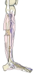
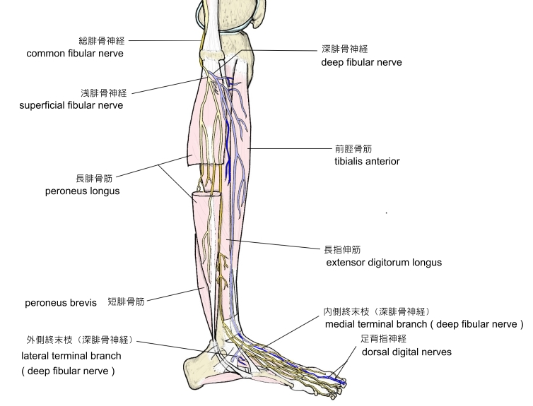
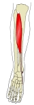
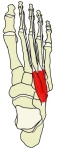
右下腿(側面)
|
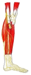
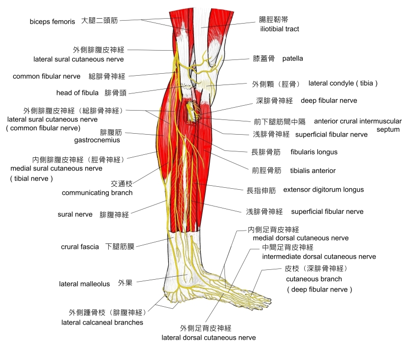
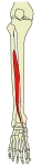
右下腿(側面)
|
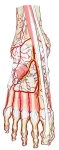
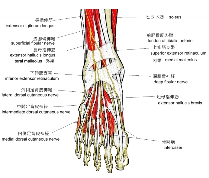
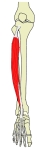
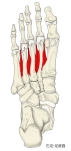
右足(背面)
|
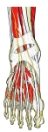
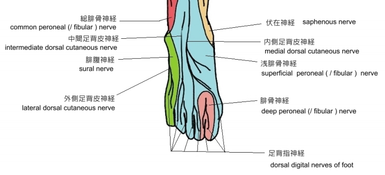
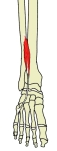
右足(背面)
|
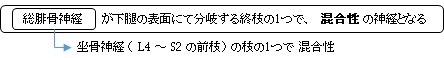
 総腓骨神経は腓骨頭を回って長腓骨筋 総腓骨神経は腓骨頭を回って長腓骨筋
以下が浅腓骨神経が総腓骨神経から分岐してからの主な走行となる。
1 . 総腓骨神経は腓骨頭を回って長腓骨筋を貫き下腿の前面に現れ浅腓骨神経と深腓骨神経に分岐する。
2 . 深腓骨神経は腓骨頭の下方で長腓骨筋/長指伸筋と前脛骨筋との間を筋枝を出しながら下行する。
3 . 最初は前脛骨動脈/静脈の外方に位置するが、下行するに従って前方に移動し、足根部においては
再び外方に位置する。
4 . 下伸筋支帯の下を通過して内側終末枝と外側終末枝に分岐する。
内側終末枝:母指方へ走行して分岐し、2本の足背指神経となる。
「 船戸和弥のホームページ 」では以下のような解説になっている。
「 内側終末は1本の背側趾神経を派生し、この神経は二分して第1、2趾の隣接面を支配する。」
外側終末枝: 短指伸筋の下を外側方に走行し、放射線状に数本の細い枝を出す。

以下は「 船戸和弥のホームページ 」を中心にして作成したものになるが、血管などはその全てを列挙できているかどうかは定かではない。

前脛骨筋 |
長母指伸筋 |
長指伸筋 |
第3腓骨筋 |
短指伸筋 |
|
短母指伸筋 |
背側骨間筋 |
浅/深腓骨動脈
|
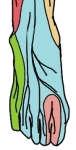
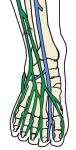
足背の皮神経
|
|

「 日本人体解剖学 (上巻) 」では「枝」として筋枝と足背指神経の2つを挙げているだけだが、「 船戸和弥のホームページ 」では、関節枝や動・静脈への枝も挙げている。よって、ここでは後者を参考に作成してみた。 」では「枝」として筋枝と足背指神経の2つを挙げているだけだが、「 船戸和弥のホームページ 」では、関節枝や動・静脈への枝も挙げている。よって、ここでは後者を参考に作成してみた。
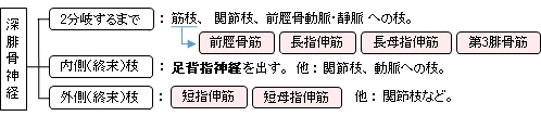
■ 2分岐するまで ■
深腓骨神経は、足首関節において内側終末枝と外側終末枝に2分岐するまでに、以下の筋に筋枝を出して支配する。
・ 前脛骨筋 ・ 長指伸筋 ・ 長母指伸筋 ・ 第3腓骨筋
また、筋枝以外に関節枝や血管への小枝があり、「 船戸和弥のホームページ 」には以下のような解説文が見られる。
「 下腿では、(途中略)足根に関節枝を送り、さらに 前脛骨動静脈 に小枝を出す。 」
■ 内側(終末)枝 ■
2分岐後に前方(母指方)に向かう神経線維で、最後には2本の背側指神経となって母指の外側縁と第2枝の内側縁の背側の皮膚に至る。また、その他の派生する神経線維として
・ 関節枝 : 近くの 中足指節関節 / 指節関節 へ行く
・ 血管へ : 足背動脈 へ行く
また、「船戸和弥のホームページ 」には以下のような解説文が見られる。
「 時として第1腓側骨間筋 に小枝を送る。」
■ 外側(終末)枝 ■
2分岐後に短指伸筋の下を外方に走り、やや放射線状に広がって数本の細い枝を出し以下に至る。
・ 筋肉へ : 短指伸筋、 短母指伸筋 また、「船戸和弥のホームページ 」には以下のような解説文が見られる。
「 時として第2および第3の 背側骨間筋 を支配する。」
・ 関節へ : 近くの 足根関節 / 足根中足関節
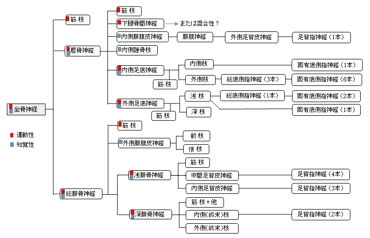
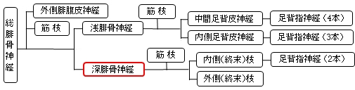
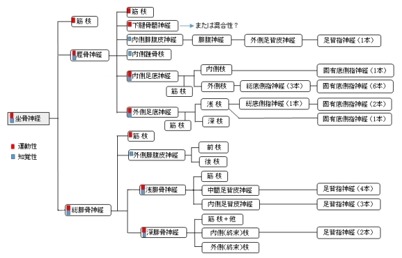
以下は「 日本人体解剖学 (上巻) 」を参考にして作成した坐骨神経の枝の簡単な図となる。

「 船戸和弥のホームページ 」では以下のように解説している。
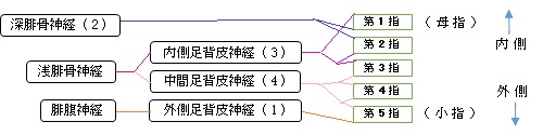
「 浅腓骨神経は総腓骨神経の終枝の一つ、腓骨筋と長趾伸筋の間を下行する。 (Netter)浅腓骨神経は、長趾伸筋と腓骨筋の間を下行し、長腓骨筋と短腓骨筋に筋枝を出した後、下腿の中部から下部1/3に移る高さで下腿筋膜を貫く。この高さで、浅腓骨神経は、内側足背皮神経と中間足背神経とに分かれる。内側足背皮神経は足根の前面を走行して足背に至り、下部下腿前面と足背の皮膚と筋膜に枝を送る。下伸筋支帯の下縁近くで、この神経は2本の足背趾神経に分岐する。このうち1本は、足背および母趾の内側面と背側面を支配し、他の1本は第2,第3趾の背側面と側面とを支配する。中間足背皮神経は、足背外側部に沿って走行し、近傍の皮膚や筋膜に枝を出し、第3と第4趾および第4と第5趾に行く2本の足背趾神経に分かれる。また、中間足背皮神経は、外側足背皮神経と交通する。 」
以下は「 Wikipedia 」の解説文となる。
「 The superficial peroneal nerve or superior fibular nerve, innervates the peroneus longus and peroneus brevis muscles and the skin over the antero-lateral aspect of the leg along with the greater part of the dorsum of the foot (with the exception of the first web space, which is innervated by the deep peroneal nerve).
【 structure 】
■ Lateral side of the leg ■
Superficial peroneal nerve is the main nerve of the lateral compartment of the leg. It begins at the lateral side of the neck of fibula, and runs through the peroneal muscles. In the middle third of the leg, it descends between the peroneus longus and peroneus brevis muscles, and then reaches the anterior border of the peroneus brevis to enter the groove between the peroneus brevis and extensor digitorum longus under the deep fascia of leg. It becomes superficial at the junction of upper two-thirds and lower one-thirds of the leg by piercing the deep fascia. Superficial peroneal nerve gives off several branches in the leg.[1]
- Muscular branches to peroneus longus and peroneus brevis[1]
- Cutaneous branches to supplies the skin over the lower one-third of the lateral side of the leg and greater part of the dorsum of the foot except for areas that are supplied by saphenous nerve(medial side of the leg), sural nerve (lateral side of the foot), deep peroneal nerve (first webbed space of the dorsum of the foot), medial and lateral plantar nerves (plantar surface of the foot).[1]
■ Foot ■
At the junction between the upper two-thirds and lower one-thirds of the leg, superficial peroneal nerve is divided into medial dorsal cutaneous nerve (medial branch) and intermediate dorsal cutaneous nerve (lateral branch).[1]
- The medial branch crosses the ankle and divides into two dorsal digital nerves - one for the medial side of the big toe, and the other for the adjoining sides of the 2nd and 3rd toes.[1]
- The lateral branch divides into two dorsal digital nerves for the adjoining sides of the third and fourth, and fourth and fifth toes.[1]
- Communicating branches - the medial branch communicates with saphenous nerve and deep peroneal nerves while the lateral branch communicates with sural nerve.[1]」
【 語 句 】
・: ・: ・: ・: ・: ・: ・: ・: ・: ・: ・: ・:
【 イラスト掲載サイト 】
・ イラストや写真を掲載しているサイト-Ⅰ
・ イラストや写真を掲載しているサイト-Ⅱ
・ イラストや写真を掲載しているサイト-Ⅲ
・ イラストや写真を掲載しているサイト-Ⅳ
・ イラストや写真を掲載しているサイト-Ⅴ

|