

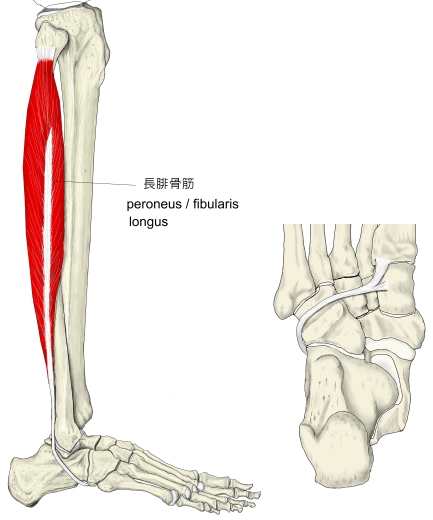
長腓骨筋 (ちょうひこつきん、英:peroneus longus muscle)
・ 概 要 |
・ 作 用 |
・ イラスト掲載サイト |
|
・ イラスト |
・ 神経 / 脈管 |
||
・ 起始 / 停止 |
・ Wikipedia |
![]()
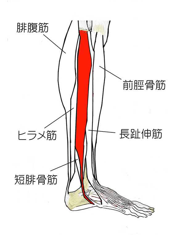
・「長腓骨筋の筋腹は、下腿部の遠位2/3の領域に存在する。そのうち、遠位1/3の領域では、筋腹は停止腱の後方のみに存在する。」(骨格筋の形と触察法)
筋連結 : 短腓骨筋、長趾伸筋、第三腓骨筋、大腿二頭筋(長頭)、長母趾屈筋、ヒラメ筋、足底筋
以下は「船戸和也のHP」の解説文となる。
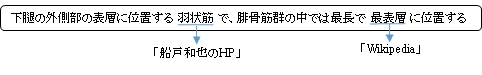
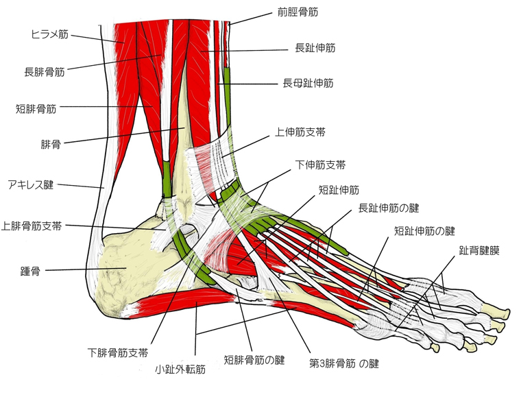
「長腓骨筋は羽状筋で、腓骨筋を容れる部を形成する壁(腓骨、筋間中隔、下腿筋膜)の近位部から起こり、第1中足骨粗面及び第2楔状骨に停止する。その腱は短腓骨筋のの筋膜と腱の上を遠位に走る。外果の後ろで長および短の腓骨筋は総腱鞘に包まれる。総腱鞘は上腓骨筋支帯によって外果に固定される。踵骨外側面で腱鞘は分かれ、短腓骨筋の腱は腓骨筋滑車の上を第5中足骨へと走る。また、長腓骨筋の腱は腓骨筋滑車のうしろを通って足の外側縁にある方向転換点へ進む。両腱は下腓骨筋支帯でしっかりと支持されている。長腓骨筋の腱は線維軟骨でおおわれた立方骨粗面上を滑走し、腱鞘に包まれて、長足底靱帯でおおわれた溝の中を通って足底を横切り、第1(2)中足骨と第二楔状骨へいたる。)」
![]()
 |
 |
 |
||||
 |
||||||
![]()
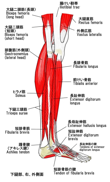
【 起 始 】
「脛骨の外側顆、腓腓関節包、腓骨の腓骨頭と外側面の近位1/3の領域および外側縁の中央1/3の領域、前下腿筋間中隔、後下腿筋間中隔、下腿筋膜」(日本人体解剖学)
「腓骨筋を容れる部を形成する壁(腓骨、筋間中隔、下腿筋膜)の近位部から起こり」(船戸和也のHP)
【 停 止 】 : 内側楔状骨(7番) ・第1中足骨の骨底(8番)(
※第2中足骨を含むこともある。)
 |
![]()
「 足を足底側に屈曲し、同時に外反する。足を固定すると下腿を後方に傾ける。」 ( 日本人体解剖学 )
![]()
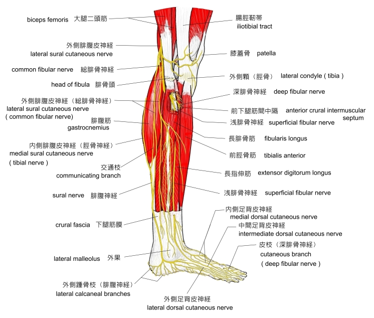
・ 神 経 : 浅腓骨神経(L5,S1)
 |
![]()
In human anatomy, the peroneus longus (also known as fibularis longus) is a superficial muscle in the lateral compartment of the leg, and acts to evert and plantarflex the ankle.
The muscle, the longest and most superficial of the three peroneus muscles, is attached proximally to the head of the fibula and its 'belly' runs down most of this bone. It becomes a tendon that goes posteriorly around the lateral malleolus of the ankle, then continues under the foot to attach to the medial cuneiform and first metatarsal. It is innervated by the superficial peroneal nerve, which arises from the fifth lumbar and first sacral roots of the spinal cord.
【Structure】
It arises from the head and upper two-thirds of the lateral surface of the body of the fibula, from the deep surface of the fascia, and from the intermuscular septa between it and the muscles on the front and back of the leg; occasionally also by a few fibers from the lateral condyle of the tibia. Between its attachments to the head and to the body of the fibula there is a gap through which the common peroneal nerve passes to the front of the leg.[2]
It ends in a long tendon, which runs behind the lateral malleolus, in a groove common to it and the tendon of the peroneus brevis; the groove is converted into a canal by the superior peroneal retinaculum, and the tendons in it are contained in a common mucous sheath.
【 語 句 】
・evert:裏返す、めくる ・peroneus muscles:腓骨筋(群) ・head of the fibula:腓骨頭 ・ lateral malleolus:外果 ・medial cuneiform:内側楔状骨 ・metatarsal:中足骨 ・superficial peroneal nerve:浅腓骨神経 ・fascia:筋膜 ・intermuscular septa:筋間中隔 ・lateral condyle:外側顆 ・tibia:脛骨 ・common peroneal nerve:総腓骨神経 ・peroneus brevis:短腓骨筋 ・superior peroneal retinaculum:上腓骨筋支帯 ・mucous:粘液を分泌する ・sheath:鞘
The tendon then extends obliquely forward across the lateral side of the foot, below the peroneal tubercle, and the tendon of the peroneus brevis, and under cover of the inferior peroneal retinaculum.
It crosses the lateral side of the cuboid, and then runs on the under surface of that bone in a groove which is converted into the peroneal canal by the long plantar ligament; the tendon then crosses the sole of the foot obliquely, and is inserted into the lateral side of the base of the first metatarsal bone and the lateral side of the medial cuneiform.
Occasionally it sends a slip to the base of the second metatarsal bone.
The tendon changes its direction at two points: first, behind the lateral malleolus; secondly, on the cuboid bone; in both of these situations the tendon is thickened, and, in the latter, a sesamoid fibrocartilage (sometimes a bone), is usually developed in its substance.
【Nerve supply】
The fibularis longus muscle is supplied by the superficial fibular nerve.
【Function 】
The peroneus longus and brevis muscles plantarflex the foot, in conjunction with the tibialis posterior, antagonizing the tibialis anterior and peroneus tertius, which are dorsiflexors of the foot.
The peroneus longus also everts the sole of the foot, and from the oblique direction of the tendon across the sole of the foot is an important agent in the maintenance of the transverse arch.
Taking their fixed points below, the peroneus muscles serve to steady the leg upon the foot.
This is especially the case in standing upon one leg, when the tendency of the superincumbent weight is to throw the leg medialward; the peroneus longus overcomes this tendency by drawing on the lateral side of the leg.
【 語 句 】
・peroneal tubercle:腓骨筋滑車 ・inferior peroneal retinaculum:下腓骨筋支帯 ・cuboid:立方骨 ・long plantar ligament:長足底靭帯 ・sesamoid:種子骨 ・fibrocartilage:線維軟骨 ・in conjunction with~:~と合わせて ・tibialis posterior:後脛骨筋 ・tibialis anterior:前脛骨筋 ・peroneus tertius:第三腓骨筋
![]()
![]()